Durgesh V1, Venugopala Rao B2*, Roja Rani CH2, and Vijaya Lakshmi K2
- Department of Anatomy, Maharajah Institute of Medical Sciences, Nellimarla, Vizianagaram District, Andhra Pradesh, India
- Department of Anatomy, Faculty of Medicine, MAHSA University, Kuala Lumpur, Malaysia.
| Corresponding Author: Venugopala Rao B, Department of Anatomy, Faculty of Medicine, MAHSA University, Kuala Lumpur, Malaysia. |
| Received: 15 September 2014 Accepted: 28 September 2015 |
Visit for more related articles at Research & Reviews: Journal of Medical and Health Sciences
The operative exposure of a fracture in an osteosynthesis causes disturbances in the blood supply, which often leads to a prolonged process of healing or even to healing problems, a fracture non-union, which is frequently located at the forearm. In order to keep the damage to the supplying vessels as little as possible, a study of the position, direction and number of the arteries of the bones of forearm is essential. In this study, we have studied the diaphyseal nutrient foramina (NF) in 144 ulnae and the findings discussed.
Keywords |
| Ulna, NF, ulnar tuberosity (UT) |
Introduction |
| Nutrient foramen is an opening through which the nutrient artery enters and supplies the shaft of the long bones. It is the main arterial supply and is particularly important during the active growth period, as well as during the early phases of ossification [1]. |
| It is described as going towards the slow growing end of the long bone which understandably requires more blood to keep pace with the opposite end of the long bone. “To the elbow I go, from the knee I flee” is a popular mnemonic among the medical fraternity. Most of the long bones have at least one NF, sometimes double and occasionally none. The position is described as somewhat in the middle of the shaft and this position is calculated by Hughes’ foraminal index. Using this index, ulna is divided into three parts [2]. Nutrient artery can be in surgical danger in cases of isolated ulnar shaft fractures (though rare but can happen while trying to fend off a blow leading to delay in fracture union). |
MATERIALS AND METHODS |
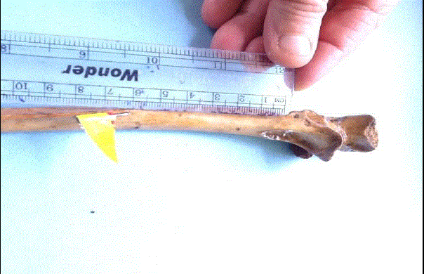
| 144 ulnae were collected from the Anatomy museum at the Maharajah Institute of Medical sciences and examined with naked eye for the NF and the number, direction and position were photographed using I-pad. The ulnae were of unknown sex. The direction and patency of the NF were examined by inserting a 24-gauge needle fitted with a coloured flag to provide a better view. The position of the NF was measured from the tip of the ulnar tuberosity by a ruler. The foraminal index was not calculated. |
RESULTS |
| 4 ulnae showed the NF on the interosseous border and one of them had two NF. These are distanced about 3 cm to 6 cm from the apex of UT. Most of the ulnae had NF on the anterior surface about 7 cm from UT. Two ulnae showed double NF on the anterior border, the proximal being 2 cm and the distal about 6 cm from UT. One ulna showed a NF on the posterior border. In one ulna, one of the NF was on the anterior border and one on the anterior surface which are 3 cm and 7 cm respectively. The entire NF were indeed directed upwards towards the elbow following the classical description. In this series, we have not come across absent NF. The distances of NF ranged from 2 cm to 7 cm from the apex of the ulnar tuberosity. None was found in the middle and distal thirds of the shaft. |
DISCUSSION |
| Many anomalies in the position were reported in tetrapods [2]. Near the elbow arteries, coming from large adjoining vessels, penetrate the area of the capsular insertion. The nutrient arteries enter both bones in the second proximal quarter of diaphysis, at the radius from anterior to medial, at the ulna from anterior to anteroradial. Small vessels, which penetrate closely proximal to the articular surface in order to supply the distal forearm bones, come from an anastomosis between the radial, the interosseous and the ulnar arteries [3]. One of the causes of delayed union or non-union of fracture is lack of arterial supply [4]. Two well-known factors may affect nutrient foramen position. These are growth rates at two ends of the shaft and bone remodelling [5]. |
| It was suggested that the pull of muscle attachments on the periosteum explained certain anomalous nutrient foramina directions [6]. |
| The ulnar artery gave off a common interosseous artery those branches into posterior and anterior interosseous vessels that course distally on the interosseous membrane. The interosseous vessels were critical for they supply the only observed vascular branches to the ulna diaphysis. The anterior interosseous vessel supplied on average 7 branches (range, 3–11 branches) to the ulna diaphysis spaced at generally regular 2-cm intervals, with the number of branches decreasing in the distal third. The posterior interosseous artery supplied an average of 11 branches (range, 9–14 branches) to the ulna diaphysis spaced at 1-cm intervals [7]. |
| Nonsurgical treatment of isolated ulnar shaft fractures (IUSF) is prone to complications and is associated with malunion and nonunion [8]. This presumably is due to vascular interruption. |
CONCLUSION |
| The present study adds to the available information about the position and number of the nutrient foramina of ulna and the surgical implications in fractures. The information may also be useful to the orthopedicians. The different positions of the NF observed might be a reflection of the phenomenon seen in lower vertebrates rather than differential growth patterns. |


