Keywords
|
| Skin Cancer, Artificial Neural Network, Segmentation, Wavelet Transform, Back Propagation. |
INTRODUCTION
|
| Skin Cancer is the cancer affecting the skin. Skin cancer may appear as malignant or benign form. Benign Melanoma is simply appearance of moles on skin. Malignant melanoma is the appearance of sores that cause bleeding. Malignant Melanoma is the deadliest form of all skin cancers. It arises from cancerous growth in pigmented skin lesion. Malignant melanoma is named after the cell from which it presumably arises, the melanocyte. If diagnosed at the right time, this disease is curable. Melanoma diagnosis is difficult and needs sampling and laboratory tests. Melanoma can spread out to all parts of the body through lymphatic system or blood. The main problem to be considered dealing with melanoma is that, the first affliction of the disease can pave the way for future ones. Laboratory sampling often causes the inflammation or even spread of lesion. So, there has always been lack of less dangerous and time-consuming methods. Computer based diagnosis can improve the speed of skin cancer diagnosis which works according to the disease symptoms. The similarities among skin lesions make the diagnosis of malignant cells a difficult task. But, there are some unique symptoms of skin cancer, such as: Asymmetry, Border irregularity, Color variation and Diameter. Those are popularly known as ABCD parameters. ABCD parameters. Asymmetry, Border irregularity, Colour, Diameter. Asymmetry is one half of the tumour does not match the other half. Border Irregularity is the unevenness of images. Colour intensity change in the lesioned region is irregular. Malignant melanoma is having a diameter greater than 6mm. |
| This paper is organized as follows: Section I gives the introduction about Skin cancer and features of skin cancers. It also gives an idea about the Computer based Skin cancer detection system. Section II describes the Automatic Skin cancer Detection system and various methods involved in the system. Section III shows the results of the Classification system. Section IV concludes the paper followed by references. |
Automatic Skin Cancer Detection System
|
| Automatic early detection system is a classification system which distinguishes Malignant Melanoma from other skin diseases. This methodology uses Digital Image Processing technique and Artificial Intelligence for the classification purpose. The input to the system is Dermoscopic Images which are in digital format. Usually such images contain noises, so they are undergone Pre-processing. In order to preserve the edges, Post-processing is done. To separate the cancerous region from healthy skin, segmentation is done. There are some unique features for the cancerous images. Those features are extracted using Two Dimensional Wavelet Transform in MATLAB software. These features are given as inputs to the Artificial Neural |
| Network based classifier. It uses Back propagation Algorithm for classification. ANN classifies Malignant Melanoma from Benign Melanoma. Thus detecting whether patient is having skin cancer or not. |
| A. Dermoscopy |
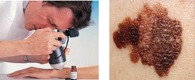
| Dermoscopy, also known as Dermatoscopy or Epiluminescence Light Microscopy (ELM). It is a kind of imaging technique used to exanimate lesions with a dermatoscope. The process is done by placing an oil immersion between the skin and the optics. Lens of a microscope is placed directly, illuminating sub-surface structures. The lighting can magnify the skin that improve on reveal most of the pigmented structure, different color shades that is not visible to naked eye; and allows direct viewing and analysis of the epidermis. The image obtained from such a dermatoscope is called Dermoscopic Image. |
| B. Image Processing |
| The Dermoscopic Image in Digital format is subjected to various Digital Image Processing Techniques. The standard image size is taken as 360x360 pixels. Usually the image consists of noises in the form of hairs, bubbles etc. These noises cause inaccuracy in classification. In order to avoid that, images are subjected to various image processing techniques. Image Processing consists of following procedures: Image Pre-processing and Post-processing. Pre-processing is done to removes the noise, fine hair and bubbles in the image. For smoothing image from noise, median filtering is used. Median filtering is a common step in image processing. Median filtering is used for minimizing the influence of small structures like thin hairs and isolated islands of pixels like small air bubbles. Post-processing is done to enhance the shape and edges of image. In addition, contrast enhancement can sharpen the image border and improve the accuracy for segmentation. |
| C. Segmentation |
| Segmentation removes the healthy skin from the image and finds the region of interest. Usually the cancer cells remains in the image after segmentation. Segmentation used is Threshold Segmentation. Thresholding often provides an easy and convenient way to perform this segmentation on the basis of the different intensities or colors in the foreground and background regions of an image. The input to a Thresholding operation is typically a grayscale or colour image. After segmentation, the output is a binary image. Segmentation is accomplished by scanning the whole image pixel by pixel and labelling each pixel as object or background according to its binarized gray level. |
| D. Feature Extraction |
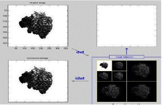
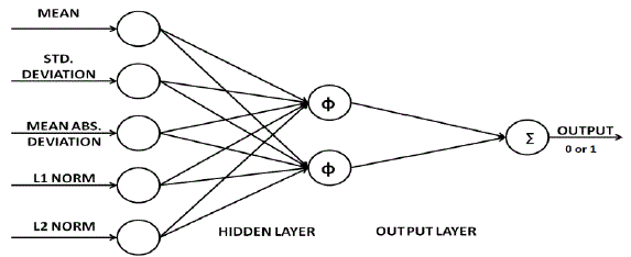
| At this stage, the important features of image data are extracted from the segmented image. By extracting features, the image data is narrow down to a set of features which can distinguish between Malignant and Benign melanoma. The extracted features should be both representatives of samples and detailed enough to be classified. 2D wavelet transform is used for the feature extraction. In this system, 2-D wavelet packet is used and the enhanced image in gray scaled as an input. Bior wavelets at two steps of decomposition are used. At each step of decomposition, the wavelet of primary image is divided into an approximate and three detailed images which show the basic information and vertical, horizontal and diagonal details, respectively. The Features extracted using the wavelet transform are: Mean, Standard deviation, Mean Absolute Deviation, L1 Norm, L2 Norm. |
| E. Artificial Neural Network Classifier |
| Classifier is used for classifying Malignant Melanoma from other skin diseases. Based on the computational simplicity Artificial Neural Network (ANN) based classifier is used. In this proposed system, a feed forward multilayer network is used. Back propagation (BPN) Algorithm is used for training. There must be input layer, at least one hidden layer and output layer. The hidden and output layer nodes adjust the weights value depending on the error in classification. In BPN the signal flow will be in feed forward direction, but the error is back propagated and weights are updated to reduce error. The modification of the weights is according to the gradient of the error curve, which points in the direction to the local minimum. Thus making it much reliable in prediction as well as classifying tasks. |
| In BPN, weights are initialized randomly at the beginning of training. There will be a desired output, for which the training is done. Supervisory learning is used here. During forward pass of the signal, according to the initial weights and activation function used, the network gives an output. That output is compared with desired output. If both are not same, an error occurs. During reverse pass, the error is back-propagated and weights of hidden and output layer are adjusted. The whole process then continues until error is zero. The network is trained with known values. After training, network can perform decision making. |
| In this proposed methodology, Five Features were given as input to a multilayer feed forward network. There is one hidden layer with two hidden neurons. Output layer with one output neuron. Activation function used is Linear function, which gives an output of 0 or 1. Zero represents non-cancerous or benign condition and one represents cancerous or malignant condition. |
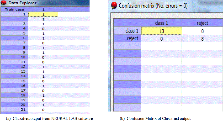
| NEURAL LAB is the software used for ANN classification. It is ANN simulation software which gives good results in classification. The network is trained using known values of Malignant and Benign Melanoma features. Many epochs of training are repeated until Mean Square Error is less than minimum value. Then data for classification is given as input to classifier. 21 Malignant and Benign Melanoma Features were given for classification. The output of the classifier is either ‘0’ or ‘1’. One represents cancerous condition and Zero represents Non-cancerous condition. |
EXPERIMENTAL RESULTS
|
| For the proposed system, 31 Dermoscopic images were collected from Internet. They were undergone Median Filtering. After that, Filtered images were segmented by Threshold Segmentation. Feature Extraction of images was done using 2D wavelet transform. All these were done in MATLAB software. Five features were selected for classification- Mean, Standard Deviation, Mean Absolute Deviation, L1 Norm, L2 Norm. The Obtained Features were given as inputs to a Feed Forward Neural Network. Activation function used is linear function, which gives an output of '0' or '1'. Zero represents non-cancerous or benign condition and one represents cancerous or malignant condition. The neural network is designed using NEURAL LAB software. The training is done with known value. After training, Data Sets for classification were given to the Network. 21 cases were given for classification. The network classifies the given data into cancerous or noncancerous. Among the 21 cases 13 were classified as cancerous and 8 non-cancerous. It is shown in the form of a Confusion Matrix as shown in Fig. 5(b). It has a good rate of accuracy too. |
| The proposed system is proved to be much convenient than the conventional Biopsy method. Since this method is Computer Based Diagnosis, there is no need for any skin removal for diagnosis. It requires only the Dermoscopic image. |
CONCLUSIONS
|
| A Computer based early skin cancer detection system is proposed. It proves to be a better diagnosis method than the conventional Bioscopy method. The diagnosing methodology uses Digital Image Processing Techniques and Artificial Neural Networks for the classification of Malignant Melanoma from other skin diseases. Dermoscopic images were collected and they are processed by various Image processing techniques. The cancerous region is separated from healthy skin by the method of segmentation. The unique features of the segmented images were extracted using 2-D Wavelet Transform. Based on the features, the images were classified as Cancerous and Non-cancerous. This methodology has got good accuracy also. By varying the Image processing techniques and Classifiers, the accuracy can be improved for this system. |
ACKNOWLEDGEMENT
|
| We would like to thank all the faculties and students of Electrical and Electronics Engineering Department, Thangal Kunju Musaliar College of Engineering management for their guidance and support and facilities extended to us. |
Tables at a glance
|
 |
| Table 1 |
|
| |
Figures at a glance
|
 |
 |
 |
 |
 |
| Figure 1 |
Figure 2 |
Figure 3 |
Figure 4 |
Figure 5 |
|
| |
References
|
- HoTak Lau and Adel Al-Jumaily, Automatically Early Detection of Skin Cancer: Study Based on Neural Network Classification, InternationalConference of Soft Computing and Pattern Recognition, IEEE , pp 375-380, 2009
- FikretErcal, AnuragChawla, William V. Stoecker, Hsi-Chieh Lee, and Randy H.Moss, Neural Network Diagnosis of Malignant Melanoma FromColor Images, IEEE Transactions on Biomedical Engineering. vol. 41, No. 9, 1994
- Ali AI-Haj, M.H.KabirWavelets Pre-Processing of Articial Neural Networks Classifiers , IEEE Transactions on Consumer Electronics, Vol. 53 Issue2, pp. 593-600 , 2008.
- PankajAgrawal, S.K.Shriwastava and S.S.Limaye, MATLAB Implementation of Image Segmentation Algorithms, IEEE Pacic Rim Conference onCommunication, Computer and Signal Processing, pp. 602-605., 2010.
- AsadollahShahbahrami, Jun Tang A Colour Image Segmentation algorithm Based on Region Growing, IEEE Trans. on Consumer ElectronicsEuromicro, pp 362-368, 2010.
- T. Tanaka, R. Yamada, M. Tanaka, K. Shimizu, M. Tanaka,A Study on the Image Diagnosis of Melanoma, IEEE Trans. on Image Processing, pp.1010-1024, June 2004.
- Y. Andreopoulos, N. D. Zervas, G. Lafruit, P. Schelkens, A local wavelet transform implementation versus an optimal row-column algorithm for the2D multilevel decomposition, IEEE International Conf. on Image Processing, volume 3, 2001.
- Mariam, A.Sheha,Mai, S.Mabrouk, AmrSharawy, Automatic Detection of Melanoma Skin Cancer using Texture Analysis, International Journal ofComputer Applications, Volume 42, 2012.
- M. Vetterli, O. Riol, Wavelets and Signal processing, IEEE Signal Processing Magazine, pp.14-38, Oct 1991.
- Shubhankar Ray and Andrew Chan, Automatic feature extraction from wavelet coefficients using Genetic Algorithms, IEEE Trans. on ImageProcessing, pp. 980-1025, June 2000.
|