ISSN ONLINE(2319-8753)PRINT(2347-6710)
ISSN ONLINE(2319-8753)PRINT(2347-6710)
J.Ramya1,S.Soundarya2,A.Nagoormeeral@Rahmathnisha3,E.Revathi4
|
| Related article at Pubmed, Scholar Google |
Visit for more related articles at International Journal of Innovative Research in Science, Engineering and Technology
Exudates are the primary signs if diabetic retinopathy which are mainly cause blindness. Diabetic retinopathy is a complication of diabetes that caused by changes in the blood vessels of the retina. It is one of the earliest and most prevalent symptoms of diseases leading to blindness such as diabetic retinopathy and wet macular degeneration. Hence, detection of exudates is an important diagnostic task. In this project, we extract the blood vessels using thresholding method and detect optic disc using circular Hough transform method. Finally we detect the exudates using adaptive thresholding method in fundus image.
Keywords |
| Diabetic retinopathies, Exudates, Fundus image, Hough transform, Macular. |
INTRODUCTION |
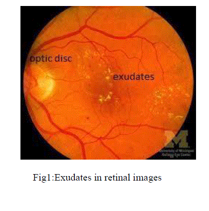
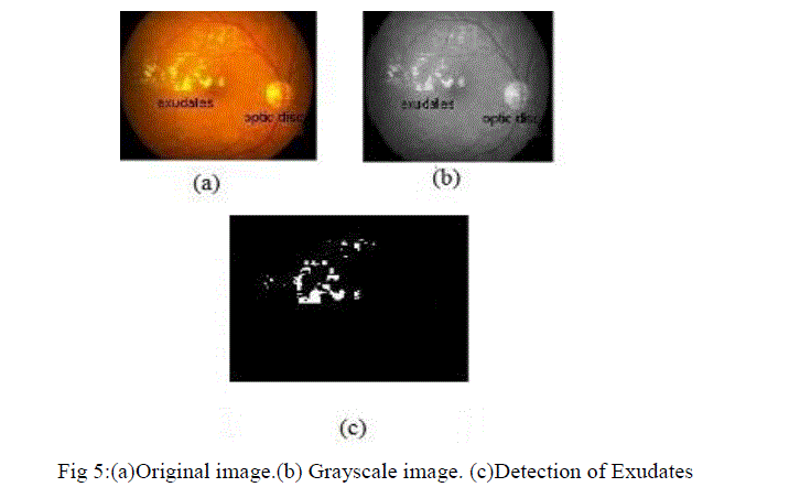
| Blindness is outcome of diabetic retinopathy. It is serve eye disease and major cause of blindness. Exudates means, blood leakage from blood vessels which is the major cause of blindness in diabetic patients and old age people. Most techniques are used to the exudates detection in fundus image. In retinal image hard exudates are appeared in various size and locations. Hard exudates are representing in yellow part seen in retina, usually posterior pole near the macula. The retinal image of eye is given in fig1, |
 |
II. RELATED WORKS |
| Region growing segmentation method use maximum variance to obtain the optic disc center and to obtain the exudates in[1]. The existing method [2] based on detecting areas of higher intensities, color and high contrast by detecting their contours. First they detect the optic disc of the image using canny edge detector. Then edge detection algorithm is used to detect their contours. Blood vessels are extracted by using morphological operations. That is based on the closing the image with two line structuring element of different sizes. They use morphological reconstruction to detect the exudates. That is work also in the lower quality retinal images. But it has low accuracy in the non-uniform illumination.[3] Using color normalization and local contrast enhancement followed by fuzzy c-means clustering and neural networks. It has low accuracy when using non uniform illumination.[4] use the blood vessel intersection property. Incorporated spatial neighborhood information into the standard FCM clustering for exudates classification is used by [5]. |
| Neural Network classifier is used to detect hard exudates using multilayer perception, radial basis functions and support vector machine [6]. To identified exudates in grey level images based on a recursive region growing technique [7]. To classify the retinal exudates Neural Network (NN) also be used in[8]. The result of classifying the whole 20X20 patches rather than a pixel resolution classification. Detection of exudates using mathematical morphology in[9].Morphological operations of different sizes are used to detect hard exudates, but there is disadvantage that is size of structuring element is suitable for one image not suitable for another image. Detection exudates using Back Propagation Neural Network in [10]. |
III.METHODS AND MATERIALS |
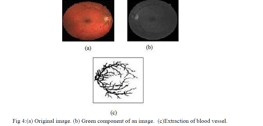
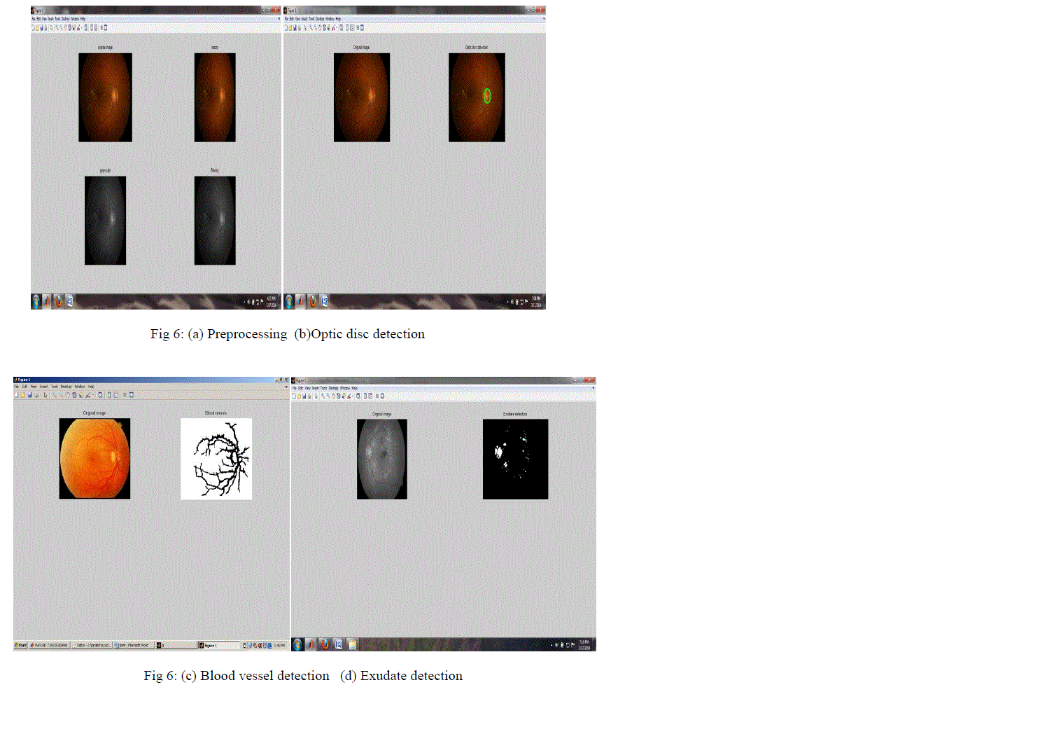
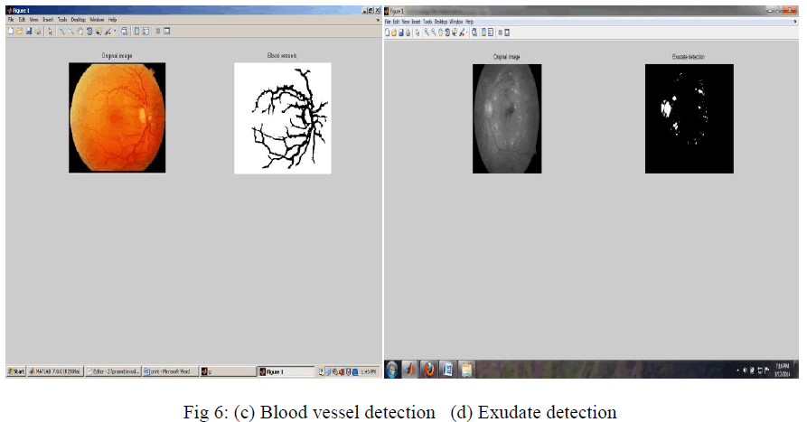
| In the proposed method, the images are collected from the DIARETDB1 [10] database that is publically available from the internet. First step is pre-processing of an image which is mainly used to remove the noise from the images. Optic disc detection is based on the circular Hough transform. Because almost all the optic disc are in circular structure. Blood vessel extractions are based on the thresholding method. After detection of optic disc and blood vessels, the detection of exudates are performed by adaptive thresholding. That is used to faint exudates also. |
A. PREPROCESSING: |
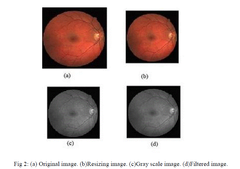
| Preprocessing images commonly involves removing low frequency background noise, normalizing the intensity of the individual particles images, removing reflections, and masking portions of images. Image preprocessing is the technique of enhancing data images prior to computational processing. Preprocessing consist of three steps, |
| ïÃâ÷ Resizing |
| ïÃâ÷ Grayscale image |
| ïÃâ÷ Filtering |
| (i)Resizing: It is used to alter the size of an image. |
| (ii)Gray scale image: It converts the rgb image to gray scale image. |
| (iii)Filtering: Filtering method used to separate the noise pixels. In our project we can use median filtering. The median filter is normally used to reduce the noise in image. Median filter works by moving through the image pixel by pixel, replacing the each value with the median value of neighbouring pixels. Median filter function is given below, |
| I = medfilt2 (img,[3 3]); âÃâÃâ¬Ã¢ÃâÃâ¬Ã¢ÃâÃâ¬Ã¢ÃâÃâ¬Ã¢ÃâÃâ¬Ã¢ÃâÃâ¬Ã¢ÃâÃâ¬Ã¢ÃâÃâ¬(1) |
| The median filter is better able to remove the outliers without reducing the sharpness of an image. It is much less sensitive than the mean to extreme values. |
 |
B.SEGMENTATION: |
| Segmentation is the part of partitioning a digital image into multiple segments. Simplify and/or change the representation of an image into something that is more meaningful and easier to analyze. Image segmentation is the front stage processing, three advantages in image segmentation that are speed, good shape and reliable. In our project image segmentation consist into two steps, |
| ïÃâç Optic disc detection |
| ïÃâç Blood vessels extraction |
(i)Optic disc detection: |
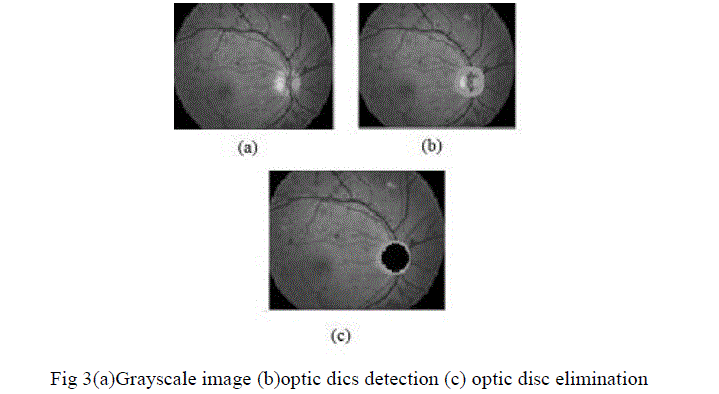
| Optic disc detection is one of the main task in detection of exudates. Because optic disc has similar characteristics of exudates such as brightness, color and contrast. Normally optic disc similar to circular structure. So we use the circular Hough transform method to detect optic disc. |
| The Hough transform are used to detect the shape of object in image. Circular transform are implemented here which is used to find the optic disc in the fundus image The advantage is that it is tolerant of gaps in feature boundary descriptions and is relatively unaffected by image noise. The circle can be represented as, |
| (x-a)²+(y-b)²+r²=0 âÃâÃâ¬Ã¢ÃâÃâ¬Ã¢ÃâÃâ¬Ã¢ÃâÃâ¬Ã¢ÃâÃâ¬Ã¢ÃâÃâ¬Ã¢ÃâÃâ¬Ã¢ÃâÃâ¬(1) |
| The two parametes x,y are the center coorddinates of the circle and another parameter r represent the radius of the circle.After the detection of optic disc we need to eliminate that. While, detection of exudates optic disc also appear on the result image that is the reason for elimination of the optic disc in Fig 3(c). |
 |
(ii) Blood vessels extraction: |
 |
| Adaptive thresholding typically takes a grayscale image or color image as an input and produce the output as binary image. For each pixel in the image, a threshold has to be calculated. If the pixel value is below the threshold it is set to the background value, otherwise it assumes the foreground value. |
 |
IV.RESULT |
| In this project we used the dataset from DIARETDB1 which is publically available. That dataset contain 89 images of resolution 1500x1152, with 47 of images contain with exudates. Remaining 42 of images contain without exudates. We used 15 images for detecting exudates. The image has been scaled to 500x500. |
| We choose sensitivity and specificity to measure the accuracy of the algorithm at the pixel level. This evaluation consider the four values such as true positive (TP), true negative (TN), false positive (FP), false negative (FN). From these quantities the sensitivity and specificity can computed as, |
 |
 |
VI.CONCLUSION |
| The color fundus images are playing a major role in the detection of exudates. Now a day, the diabetic patients are increased throughout the world. So early detection of exudates are useful to reduce the vision loss occurs. At the same time automatic detection of exudates are useful to the ophthalmologists. The main purpose of the project is to detect hard exudates. The proposed algorithm is also work in lower quality retinal images. |
VII.FUTURE WORK |
| Future work will be the detection Microneurysm and Haemorrhage could be added to the system, so that it increases the ability to verify the diabetic retinopathy. There may be problem in separating the pathologies from small vessels. It could be useful to extent this work to detect them. |
References |
|