ISSN ONLINE(2319-8753)PRINT(2347-6710)
ISSN ONLINE(2319-8753)PRINT(2347-6710)
Dr. Shweta Mishra1, Dr. Anita Munde2, Dr.Safia Shoeb3, Dr. Sunil Sahuji4, Dr. Pranali Wankhede5
|
| Related article at Pubmed, Scholar Google |
Visit for more related articles at International Journal of Innovative Research in Science, Engineering and Technology
Keywords |
| erupted, odontoma, odontogenic tumor. |
I.INTRODUCTION |
| The term odontome by definition alone refers to a tumor of odontogenic origin consisting of mesenchymal and epithelial dental elements. Histologically, they are composed of different dental tissues, including enamel, dentine, cementum and, in some cases, pulp tissue.2,3 |
| According to WHO classification odontomes can be divided into three groups. |
| Complex odontome: When the calcified dental tissues are simply arranged in an irregular mass bearing no morphologic similarity to rudimentary teeth. |
| Compound odontome: Composed of all odontogenic tissues in an orderly pattern that result in many teethlike structures, but without morphologic resemblance to normal teeth. |
| Ameloblastic fibro-odontome: Consists of varying amounts of calcified dental tissue and dental papilla-like tissue, the later component resembling an ameloblastic fibroma. The ameloblastic fibro-odontome is considered as an immature precursor of complex odontome.4 |
| When a comparision is made between the occurrence rate of compound and complex odontome then usually the former is twice as common than the latter.2 |
| The purpose of this report is to add on to the rare entity of erupted complex odontome in the existing literature and thereby discuss the clinical, radiological and histopathological evidence which supported this case. |
II. CASE REPORT |
| A 20-year-old female reported to the Department of Oral Medicine and Radiology with the chief complaint of pain in upper left posterior region of the jaw since two days. Pain was spontaneous in onset, dull, intermittent, nonradiating, aggravates on chewing and relieved by itself after few minutes. Extraoral examination was unremarkable. Intraoral examination revealed a partially erupted yellowish colored hard mass of approximate size 1.5 x 2 cm on the alveolar ridge distal to left maxillary first molar. Maxillary second and third molar were missing. The surrounding mucosa was noninflamed and there was no evidence of pus discharge or draining sinus with the same. On palpation the mass was bony hard in consistency and there was no expansion with either buccal or lingual cortical plate (Fig 1 ). |
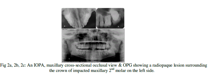
| Radiographic examination (Iopa, maxillary occusal and panoramic) ( Fig 2a,2b,2c ) revealed an impacted maxillary 2nd molar on the left side with an ill defined radiopacity with slightly greater density than the bone present with the coronal aspect of the same. Internal structure revealed radiopacity with trabecular pattern. Maxillary third molar was missing. The radiopaque mass was surrounded by a radioulucent halo which in turn had a well corticated border outside. |
| Considering the clinical and radiographic presentations, a diagnosis of erupted complex odontomas was determined. Under local anesthesia the access to the mass was achieved through an intraoral approach and odontome was extracted (Fig 3a,3b) and subjected to histopathological examination which confirmed the diagnosis of complex odontoma. Patient was kept under observation. Post operative follow up was done after 6 months which showed normal eruption pattern of left maxillary 2nd molar teeth. ( Fig 4a,4b). |
III. DISCUSSION |
| The term “Odontoma” was coined by Paul Broca in 1867.1 It is the commonest hamartoma and odontogenic tumor which constitutes for 22 % of all odontogenic tumors of the jaw. |
| The WHO classification defines Complex odontome as “a malformation in which all the dental tissues are represented, individual tissues being mainly well formed but occurring in a more or less disorderly pattern ” 5,6 as well as compound odontome which consists of multiple well formed teeth or denticles. |
| There are many etiologies of odontome formation but mostly odontomas have been associated with trauma during primary dentition, inflammatory and infectious processes, hereditary anomalies (Gardner syndrome, Hermann's syndrome).7 It has slight predilection in males with M : F ratio varies between 1.5 : 1 and 1.6 : 1. Mainly odontomas in anterior segment of jaw are compound composite in type (61%) whereas the majority in posterior segment are complex composite type. 5 Many authors have given varied types of odontomas like compound , complex, Dilated, hybrid, Geminated & Cystic.4,8,3,9 Odontomas are also classified as intraosseous and extraosseous odontomas. Erupted odontomas are rare and quite infrequent in literature.4 |
| The first case of erupted odontoma was deciphered by Rumel et al in 1980. In the year 2009 Gabriel Serra Serra et al described three cases before which only 17 cases of erupted odontomas were reported in the literature. These concluded almost 20 cases altogether.2 In 2010 Avinash et al. and Chandra et al. came up with another two cases of erupted odontomas.7,4 Further in 2011 Nisha et al documented one more case followed by another case by Dawasaz Ali zahar et al in 2012. 1,5 Out of previous 24 cases, 13 cases were of complex odontome and 11 cases were of compound odontome . Also out these 24 cases , 9 were male and 14 were female and in one case no age and sex were presented. The mean age of the patient was 20-25 yrs. We here have a rare case of erupted complex odontome which was erupted in the left posterior maxilla. |
| These are usually asymptomatic lesions, but sometimes their eruption in the oral cavity may give rise to pain, inflammation and infection as well as they are associated to alterations in permanent or temporary tooth eruption.2 Impaction and/or altered eruption occurs because the odontoma obstructs the eruptive trajectory of the teeth. Our case here presented with pain and roughness with the affected area. |
| The mechanism of odontoma eruption appears to be different from tooth eruption because of the lack of periodontal ligament and root in odontoma. The force required to move the teeth is the contractilitity of the fibroblasts,which is absent in case of odontomas. Although there is no root formation in odontoma, its increasing size may lead to the sequestration of the overlying bone and, hence occlusal movement or eruption.with increasing size over time produces a force sufficient to cause bone resorption. 1 |
| Differential diagnosis may include other opaque jaw lesions like cement-ossifying fibroma, osteoma etc . 10,8 The treatment protocol comprises of surgical extraction, fenestration & posterior orthodontic traction or simple observation with periodic clinical and radiological controls, to evaluate the course of these teeth. In our patient surgical extraction was performed and patient was kept under observation for evaluation of the normal eruption of the 2nd maxillary molar in the oral cavity. |
 |
 |
IV. CONCLUSION |
| This paper presented case of erupted complex odontoma which is rare in nature as odontomas infrequently erupt in the oral cavity. The extraction was carried out with the odontoma and patient was kept under obnservation for evaluation of the affected tooth. |
References |
|