ISSN ONLINE(2319-8753)PRINT(2347-6710)
ISSN ONLINE(2319-8753)PRINT(2347-6710)
Dr Jayant Ambulgekar1, Dr Rizwan Sanadi2, Dr Manan Doshi3 Dr Subodh Gaikwad4
|
| Related article at Pubmed, Scholar Google |
Visit for more related articles at International Journal of Innovative Research in Science, Engineering and Technology
If restorative dentistry is performed on teeth where the periodontal prognosis is not determined & disease not treated, the loss of restored tooth can occur. Restorative dentistry must be performed on a periodontium free of inflammation for a good functional & aesthetics restorative result. Surgical crown-lengthening procedures are performed to provide retention form to allow for proper tooth preparation, impression procedures & placement of restorative margins & to adjust gingival levels for aesthetics. It is important that crown-lengthening surgery is done in such a manner that the biologic width is preserved. The present case report demonstrates that surgical crown lengthening is one of the most important surgical techniques in full mouth rehabilitation of mutilated dentition.
Keywords |
| Crown lengthening, biologic width, mutilated dentition. |
INTRODUCTION |
| The surgical procedure to expose adequate clinical crown to prevent the placement of the crown margin into the area of the biologic width is termed crown lengthening surgery. Surgical crown-lengthening procedures are performed to provide retention form to allow for proper tooth preparation, impression procedures, [1]& placement of restorative margins & to adjust gingival levels for aesthetics.[2],[3] A gingivectomy technique can be used to eliminate the tissues that form the pocket or sulcus wall; such tissue may be overgrown (gingival pocket) & may interfere with the intended restorative procedures. This technique does not lengthen the clinical crown & therefore is not considered true crown lengthening surgery. By definition, the clinical crown is that portion of the tooth coronal to the alveolar crest. Therefore the bone margin must be removed to lengthen it. This is accomplished with an apically positioned flap & ostectomy, which means that tooth-supporting bone is removed. The biologic width is defined as the physiologic dimension of the junctional epithelium & connective tissue attachment. Studies have indicated that the average lengths of the connective tissue attachment & junctional epithelium are 1.07 & 0.97 mm, respectively. [4] Therefore average length of the biologic width is about 2 mm. If the restorative margin is placed into this area, the crestal bone will be lost to re-establish the biologic width. Thus, it is essential that there be at least 3 mm between the most apical extensions of the restoration margin & the alveolar bone crest. |
CASE REPORT & SURGICAL PROCEDURE |
A. Case report |
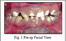
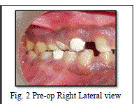
| 56 years male patient was reported to the Dept. of Periodontics, YMT Dental College & Hospital, Navi Mumbai with the chief complaint of generalized sensitivity to hot & cold since last 4-5 months. No abnormality was detected on extra-oral examination. On intra-oral examination, generalized attrition was seen with reduced clinical crown length & collapsed bite. Thus, the patient was 1st referred to Dept. of Endodontics for full mouth root canal treatment & then to the Dept. of Prosthodontics for full mouth rehabilitation. Hence, full mouth crown lengthening after full mouth root canal was selected as treatment of choice followed by prosthetic rehabilitation.(Figures 1-11) |
B. Surgical Procedure |
| Infiltration of local anaesthesia ( 2% lignocaine with 1:80,000 adrenaline) |
| Evaluation of supra osseous gingival dimensions |
| Bleeding points were marked on buccal & lingual surfaces to expose desired amount of crown |
| Inverse bevel incision following bleeding points was made by preserving as much keratinized gingiva as possible & developing interproximal papilla to facilitate wound closure |
| Crevicular incision to remove tissue wedge |
| Mucoperiosteal flap was elevated facially as well as lingually |
| Osseous resection was performed to achieve 3 mm distance between the most apical extensions of the restoration margin & the alveolar bone crest. |
| Flap was repositioned & sutures (3-0) were placed |
| Analgesics & antibiotics were prescribed to the patient & recalled after 1 week |
 |
 |
DISCUSSION |
| The dimension of space that the healthy gingival tissues occupy above the alveolar bone is now identified as the biologic width. Most authors credit Gargiulo, Wentz & Orban’s study on cadavers with establishing the dimensions of the space required by the gingival tissues. [4] Restorative considerations frequently dictate the placement of restorative margins beneath the gingival tissue crest. Restorations may need to be extended gingivally to create adequate resistance & retention form in the preparation, to make significant contour alterations because of caries or other tooth deficiencies, or to mask the tooth/restoration interface by locating it subgingivally. When the restoration margin is placed too far below the gingival tissue crest, it impinges on the gingival attachment apparatus & creates a violation of biologic width. [5] In crown lengthening procedure, gingival recession is a potential risk after removal of bone. [6] It has been found that highly scalloped thin gingiva is more prone to recession than a flat periodontium with thick fibrous tissue. [7] Also, it has been theorized that infringement on the biologic width by the placement of restoration within its zone may result in gingival inflammation, [8] pocket formation & alveolar bone loss. The methods of surgical clinical tooth crown restorations include gingivectomy, apically positioned flap and apically positioned flap with ostectomy and osteoplasty. Indications of gingivectomy and apically positioned flap for clinical tooth crown lengthening are limited and is mainly used in case of gummy smile when because of excess of gingiva, anatomical tooth crown is opened partially but gingival biological width is not altered [9]. The main method performing surgical clinical tooth crown lengthening is apically positioned flap with bone reduction which was chosen in the present case, by which biologic width can be altered to attain a 3 mm of distance between gingival margin and bone crest. |
| Other important clinical parameters that should be considered during crown lengthening is amount of attached gingiva. It has been shown that to maintain periodontal health there should be 2-3mm of attached gingiva [10]. In the present case scalloped incision was given by preserving as much of attached gingiva as possible. |
CONCLUSION |
| Good communication between the restoring dentist & the periodontist is important to achieve optimal results with crown-lengthening surgery, particularly in esthetically demanding cases. In addition to establishing the smile line, the restoring dentist evaluates the anterior and posterior occlusal planes for harmony and balance, as well as the anterior and posterior gingival contours. Surgical crown lengthening can be used to improve collapsed vertical dimension as well as collapsed bite. |
References |
|