Anil Kumar1*, and Mahindra Nagar2
- Department of Human Structure and Neurobiology, Oman medical college affiliated with West Virginia University (USA), Sohar, Sultanate of Oman
- Department of Anatomy, University College of Medical Sciences & Guru Teg Bahadur Hospital, Delhi, India
| Corresponding Author: Anil Kumar, Department of Human Structure and Neurobiology, Oman medical college affiliated with West Virginia University (USA), Sohar, Sultanate of Oman |
| Received: 01 September 2014 Accepted: 24 September 2015 |
Visit for more related articles at Research & Reviews: Journal of Medical and Health Sciences
This study proposed to morphologically analyze the adult human occipital condyle, estimate the bilateral differences, sexual dimorphism and compare with the available data. We conducted this study on hundred occipital condyles in fifty dry human skulls. The measurement was performed by the means of the vernier’s caliper on both sides of the skull. The parameters measured are length, width, height, anterior and posterior intercondylar distance of the occipital condyles. The data is tabulated and statistically analyzed. The mean length, width and the height of the left side of the occipital condyles are higher compare to the right side. The sexual dimorphism is also seen in this study. The male occipital condyles are comparatively bigger than the female one. Also the anterior and posterior intercondylar distance in male is comparatively higher. The use of morphometric values of the occipital condyles in North Indian skeletal populations may be considered in cases of fragmented cranial bases when no other morphogenetic or morphometric method can be utilized for sex determination. The data of the present study may provide anatomical reference to the neurosurgeons and thus help in surgical procedures around the craniovertebral junction
Keywords |
| Occipital condyle, Sexual dimorphism, anterior intercondylar distance, posterior intercondylar distance, Craniovertebral junction |
Introduction |
| The measurements of skeletal bones mainly neuro-cranium and viscero-cranium are often used for human population morphological studies of Age estimation, Sex determination, Stature, Ethnicity which are relevant aspects of forensic investigations and anthropological examination of unknown individuals. The human occipital condyles are unique bony structures connecting the cranium and the vertebral column. It is the only articulation between the occipital bone and the atlas hence an important part of the cranio-vertebral junction [1,2]. The integrity of occipital condyles is of vital importance for the stability of the cranio-vertebral junction. In the last two decades, the cranio-vertebral junction has been the focus of a variety of anatomical and biomechanical studies [3,4]. |
| Today, the newer neuro-imaging techniques have increased the interest for aggressive cranio-vertebral surgeries. Space occupying lesions ventral to the spinal cord at the level of foramen magnum can be surgically reached using a ventral or dorsal approach. The ventral approach has a lot of difficulties and a high rate of morbidity therefore necessitating the increasing trends of dorsal and lateral trans-condylar approach which requires partial or complete resection of the occipital condyles for access to the ventral and ventro-lateral areas of the foramen magnum [3,5,6]. Therefore knowing the anatomy and analyzing the morphometric aspects of occipital condyles is extremely important as it will help the neurosurgeon in the planning of surgical intervention involving the skull base safe and easier due to the increasing trends for transcondylar approach. The aim of the present study is an attempt to Morphometrically analyzes the adult human occipital condyles, estimate the sexual dimorphism and compare our data with the available data. |
MATERIAL AND METHODS |
| The study was conducted on hundred occipital condyles in fifty dry (50) adult human skulls obtained from the teaching skeletal collections at the Anatomy Department of University College of Medical Sciences and Guru Teg Bahadur Hospital, Delhi. All bones which showed a history of trauma or evidence of dismorphosis were excluded |
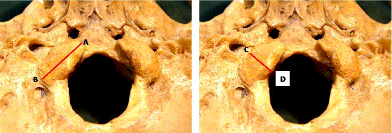
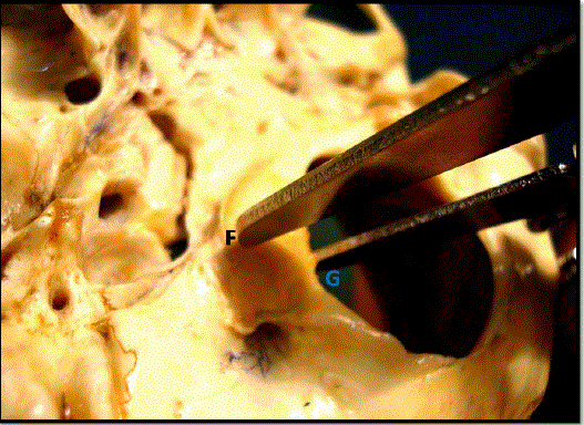
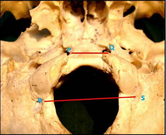
| The sex was determined by considering the classic anatomical characteristics. [7,8] All parameters were measured independently by two different observers, with a predetermined methodology to prevent inter-observer and intra-observer error. Measurements were performed by means of vernier calipers accurate to 0.1mm on occipital condyles of both sides. The parameters measured are the length or antero-posterior axis of the occipital condyles which is defined as the longest antero-posterior axis from the anterior end to the posterior end of the condyle. The width or transverse axis of the occipital condyles is the largest diameter from the medial to the lateral border of the condyle (Fig 1). The Centre or midpoint of the occipital condyle was taken as the transection of the midpoint of antero-posterior and transverse axis and the height was measured on the centre of the condyle (Fig 2). Anterior inter-condylar distance is the distance between anterior tips of right and left occipital condyles. Posterior inter-condylar distance is the distance between posterior tips of right and left occipital condyles (Fig 3). The data of these parameters are collected and tabulated. The statistical analysis was performed using SPSS 11.5 software. P < 0.001 was considered as a significant level. |
RESULTS AND DISCUSSION |
| The mean length of the right side of male and female occipital condyle was 23.88 ± 1.5 and 22.6 ± 1.30 whereas on the left side was 24.99 ± 1.82 and 24.20 ± 1.62 respectively. The mean width of the right side of male and female occipital condyle was 12.97 ± 1.43 and 1.65 ± 1.33 whereas on the left side was 14.11 ± 1.01 and 13.85 ± 1.02 respectively. The measured length of the condyles is in conformity with the length reported by Naderi, Guidotti, Oliver and Bozbuga but much larger as compared to those by Lang [9,10,11,12,13]. The average length and width of the left side compare to the right side is statistical significantly high. Also the average length and width of the male occipital condyle compare to the female is statistical significantly high. However, the statistically significant difference between the right and left axial lengths of our study disagree with those obtained by Lang [13], probably the difference in opinion is due racial variations in the cranial morphometric and the difference in methodology (Table 1). The mean height of the right side of male and female occipital condyle was 08.64 ± 0.74 and 06.92 ± 0.72 whereas on the left side was 09.32 ± 0.78 and 9.21 ± 0.76 respectively. The average height of the left side compare to the right side is statistical significantly high. Also the average height of the male occipital condyle compare to the female is statistical significantly high. The mean height of the occipital condyles is smaller than the height reported by Naderi et al [9] but in conformity with Oliver [11]. The differences between the results are possibly due to racial variations in the cranial morphometric (Table 2). Naderi et al [9] measured it in Turkish populations. The axial length and width of the left and right occipital condyles have been found to be significantly greater in males therefore sexual variation is present (Table 3). This is probably due to the presence of inter-correlation and structural relationships between the antero-posterior axis on both sides and the diameters (sagittal and transverse) of the foramen magnum which have been reported to be significantly greater in males than in females [14]. The dimensions of the foramen magnum and the axial and antero-posterior lengths of the occipital condyles are very important factors |
| Naderi et al [9] classified occipital condyles according to the length as follows: |
| • Type 1 (short): Condyles shorter than 20mm. |
| • Type 2 (moderate): Condyles between 20–26 mm. |
| • Type 3 (long): Condyles longer than 26mm. |
| Based on this Naderi et al [9] classification, the occipital condyles in the present study may be classified as type-II (moderate) variety. |
| The mean anterior and posterior intercondylar distance of occipital condyle was 17.63 mm and 42.02 mm respectively. The statistical significant difference between males and females in the anterior intercondylar distance is in conformity with Aynur Emine study [15], which he considered as another possible explanation of the inter-correlation between the significantly greater dimensions of the foramen magnum in males and the significantly wider anterior intercondylar distance (Fig. 3). According to Lang [13] the mean anterior intercondylar distance was 23.6 mm. In our study, the mean was 17.30 mm in females and 17.63 mm in males (Tables 4). The lower values may be due to the difference in methodology. We measured the distance between the anterior inner points of the occipital condyles (Fig. 3), while Lang measured the distance between the anterior circumferences of the condyles [13]. |
CONCLUSION |
| The mean antero-posterior axis, transverse axis, and height of the occipital condyles are greater on the left side as compare to the right in both sexes (Table 1). The mean antero-posterior axis, transverse axis, and height of the occipital condyles are greater in males as compare to the females (Table 3). The anterior and posterior intercondylar distance is more in males as compare to females (Table 4) and less as compare to other races (Turkish, Malawian, Caucasian, Brazilian, Chinese, and English populations). Results indicate that expression of sexual dimorphism in the occipital condylar region within the Indian population is demonstrable. Therefore, the use of morphometric values of the occipital condyles in North Indian skeletal populations may be considered in cases of fragmented cranial bases when no other morphogenetic or morphometric method can be utilized for sex determination. |
ACKNOWLEDGEMENT |
| I would like to express my profound gratitude to Dr. Mahindra Nagar and Dr. Veena Bharihoke who enlightened my period of work and invaluable guidance and continuous inspiration. Also like to thanks Mr. Laxman Singh for their assistance and technical support. |
References |
- Iscan, MY. Global forensic anthropology in the 21st century. Forensic Sci Int. 2001;117:1-6.
- Harvati K, Weaver TD. Human cranial anatomy and the differential preservation of population history and climate signatures. Anat Rec A DiscovMol Cell Evol Biol. 2006; 288 :1225-33.
- Al-Mefty O, Borba LA, Aoki N, Angtuaco E, Pait TG. The transcondylar approach to extradural nonneoplastic lesions of the craniovertebral junction. J Neurosurg. 1996;84:1âÃâ¬Ãâ6.
- Ziyal MI, Salas E, Sekhar LN. The surgical anatomy of six variations of extreme lateral approach. Turkish Neurosurg. 1999;9:105âÃâ¬Ãâ12.
- Wen HT, Rhoton AL, Katsuta T, de Oliveira E. Microsurgical anatomy of the transcondylar, supracondylar and paracondylar extensions of the far-lateral approach. J Neurosurg. 1997;87:555âÃâ¬Ãâ85.
- Sen CN, Sekar LN. Surgical management of anteriorly placed lesions at the craniocervical junction: an alternative approach. ActaNeurochir (Wien). 1991;108:70.
- Testut L, Latarjet A. Tratado de anatomiahumana, Salvat. Barcelona. 1977.
- Gray H. Caracteristicascraneales en las diferentesedades; in Williams and Warwick, Anatomia, Salvat. Barcelona. 1985.
- Naderi S, Korman E, Citak G, GÃÆüvenÃÆçer M, Arman C, SenoÃâßlu M, Tetik S, Arda MN. Morphometric analysis of human occipital condyle. ClinNeurolNeurosurg. 2005;107(3):191-9
- Guidotti A. Morphometrical considerations on occipital condyles. AnthropolAnz 1984;42:117âÃâ¬Ãâ9.
- Oliver G. Biometry of the human occipital bone. J Anat. 1975;120:507âÃâ¬Ãâ18.
- Bozbuga M, Ozturk A, Bayraktar B, Ari Z, SÃâø ahinoglu K, Polat G et al. Surgical anatomy and morphometric analysis of the occipital condyles and foramen magnum. Okajimas Folia AnatJpn. 1999;75:329âÃâ¬Ãâ34.
- Lang J, Hornung G. The hypoglossal channel and its contents in the posterolateral access to the petroclival area. Neurochirurgia. 1993;36:75âÃâ¬Ãâ80.
- Catalina-Herrera CJ. Study of the Anatomic Metric Values of the Foramen Magnum and Its Relation to Sex. Acta Anat. 130: 344-7, 1987.
- AynurEmine ,CicekciBasi, Khalil AwadhMurshed, TanerZuylan, MuzafferSeker, IsikTuncer. A Morphometric Evaluation of Some Important Bony Landmarks on the Skull Base Related to Sexes. Turk J Med Sci. 2004;34:37-42.
|


