ISSN ONLINE(2278-8875) PRINT (2320-3765)
ISSN ONLINE(2278-8875) PRINT (2320-3765)
Nikhil Amrutkar1, Yogesh Bandgar2, Sharad Chitalkar3, S.L.Tade4
|
| Related article at Pubmed, Scholar Google |
Visit for more related articles at International Journal of Advanced Research in Electrical, Electronics and Instrumentation Engineering
The primary cause of blindness in India and worldwide is recognized as Diabetic Retinopathy, which is an eye disorder caused by diabetes.For diagnosis of diabetic retinopathy, ophthalmologists use retinal image of a patient which is known as fundus image. This fundus image is captured using digital camera known as opthalmoscope.This paper presents an automated image processing system which detects the abnormality as Proliferative diabetic retinopathy(PDR) using image substraction technique and Severe diabetic retinopathy(SDR) using blob detection technique. It also gives the severity in percentage from which we get an idea that a patient is on the verge of diabetic retinopathy or not which further helps to avoid blindness
Keywords |
| Diabetic Retinopathy, Image processing,fundus image,blob detection, image substraction,SDR,PDR. |
INTRODUCTION |
| The effect of diabetes on the eye is called Diabetic Retinopathy (DR) which can lead to partial or even complete loss of vision if left undiagnosed at the initial stage. Diabetic Retinopathy is the leading cause of blindness in the working age population of developed countries. That is the reason for which efforts that has been undertaken in last few years in developing tools to assist diagnosis of diabetic retinopathy. DR is caused by changes in the blood vessels of the retina. In some people with DR, blood vessels may swell and leak fluid or abnormal new blood vessels grow on the surface of the retina. Images of patient with DR, can exhibits red and yellow spots which are problematic areas indicative of haemorrhages and exudates. |
| DR has mainly four stages: |
| A) Mild Non-Proliferative Retinopathy- At this early stage, micro-neurysms may occur.These manifestations of the disease are small areas of balloon-like swelling in the retinas tiny blood vessels. Approximately 40 percent of people with diabetes have at least mild signs of DR. |
| B) Moderate Non-Proliferative Retinopathy- As the disease progresses,some blood vessels that nourish the retina are blocked. Cotton wool spots and limited amount of venous bleeding can be seen. Generally 16 percent of patient with moderate NPDR will develop PDR within one year. |
| C) Severe Non-Proliferative Retinopathy- Many more blood vessels are blocked, depriving several areas of the retina with their blood supply. These areas of the retina send signals to the body to grow new blood vessels for nourishment. |
| D) Proliferative Retinopathy-This is the advanced stage,the signals send by the retina for nourishment trigger the growth of new blood vessels. These new blood vessels are abnormal and fragile.They grow along the retina and along the surface of the clear,vitreous gel that fills the inside of the eye. By themselves,these blood vessels do not cause symptoms or vision loss. However,they have thin, fragile walls.If they leak blood,severe vision loss and even blindness can result. About 3 percent of people in this condition may suffer severe visual loss. |
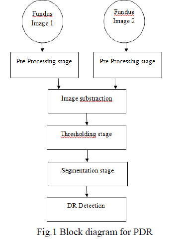
| Segmentation of blood vessels in retinal images allows early diagnosis of disease;automating this process provides several benefits including minimizing subjectivity and eliminating a painstaking,tedious task. Previous approaches,while satisfactory in some cases,still leave room for improvement, especially in abnormal retinal images. We propose to utilise blob detection and image substraction algorithm for detection of proliferative and severe diabetic retinopathy. |
BLOCK DIAGRAM |
A. Proliferative Diabetic Retinopathy detection |
 |
B. Severe Diabetic Retinopathy detection |
 |
MATERIALS AND METHODOLOGY |
A.Image Acquisition |
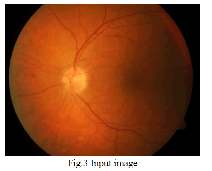
| To evaluate the performance of this method,the digital retinal images were acquired using digital camera known as opthalmoscope.Fig(3) shows the input image. Fundus image is the interior surface of the eye,opposite the lens,and includes the retina,optic disc,macula,blood vessels and fovea.We tested and evaluated our proposed algorithm on several fundus images.The image set contains both normal and abnormal cases. |
 |
B.Pre-processing |
| Pre-processing stage can be regarded as the bedrock of this work.The aim of pre-processing is to attenuate the noise,to improve the contrast and to correct the non-uniform illumination. Pre-processing mainly includes following stages: |
1)Intensity conversion: |
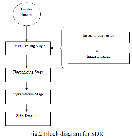
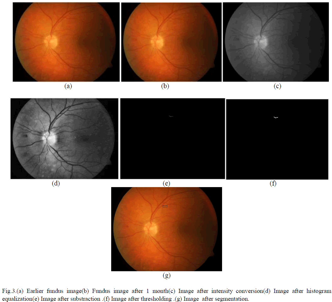
| Fig.3(c) and Fig 4(b) shows the result of intensity conversion. In digital image processing,images are either indexed images or RGB(Red,Green,Blue) images.In the RGB images the green channel exhibits the best contrast between the vessels and background while the red and blue ones tends to be more noisy.Hence intensity conversion of image is done using green channel,as the retinal blood vessels appears darker in gray image. |
2)Filtering: |
| Filtering is used to suppress the unwanted noise which gets added into the fundus image.Here median filtering is used as it is very robust and has the capability to filter any outliers and is an excellent choice for removal of salt and pepper noise. |
3)Adaptive Histogram Equalization: |
| Fig.3(d) shows the result of adaptive histogram equalization.Histogram equalization is performed to improve the image quality.Histogram equalization is nothing but a finding of cumulative distribution function for a given probability density function.After the transformation,the image will have an increased dynamic range,high contrast and probability density function of the output will be uniform.Instead of using normal histogram equalization, adaptive histogram equalization is used as it operates on small regions in the image which are called tiles.Adaptive histogram combines neighbouring tiles using bilinear interpolation to eliminate artificially induced boundaries. |
C.Thresholding |
| Fig.3(f) and Fig 4(c) shows the result of image thresholding.Thresholding is method of segmenting image based on the pixel intensity value.Thresholding is used to convert an intensity image to a binary image.Otsu’s method is used to automatically perform histogram shape-based image thresholding.Otsu’s method chooses the threshold to minimize the interclass variance of the black and white pixels. |
D.Labeling |
| Fig.4(d) shows the result of labelling. Connected components are labelled by scanning an image pixel by pixel in order to identify connected pixel regions.It groups its pixels into components based on pixels connectivity.After group formation each group is labelled with different colour. |
E.Segmentation |
| Fig 3(g) and Fig.4(e) shows the result of segmentation.The main objective of segmentation is to group the image into regions with same characteristics.The goal of the segmentation is to simplify and /or change the representation of an image into something that is more meaningful and easier to analyze.Image segmentation is typically used to locate objects and boundaries(lines,curves etc.)in the images.The result of image segmentation is a set of segments that collectively cover the entire image,or a set of contours extracted from the image. |
| After performing all above operations on the fundus image blobs are detected which is the sign of severe diabetic retinopathy. Also for the mild severity,we used image substraction algorithm in which earlier and present fundus image is substracted to detect the new increased blood vessels. |
RESULTS |
A. Results for PDR detection |
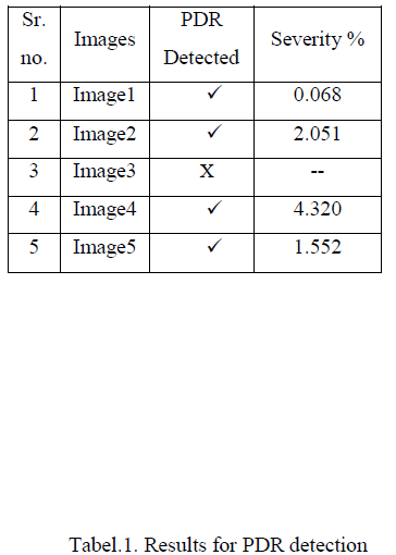
| For PDR detection,same pre-processing operations are performed on both earlier and present image. |
 |
B.Results of SDR detection |
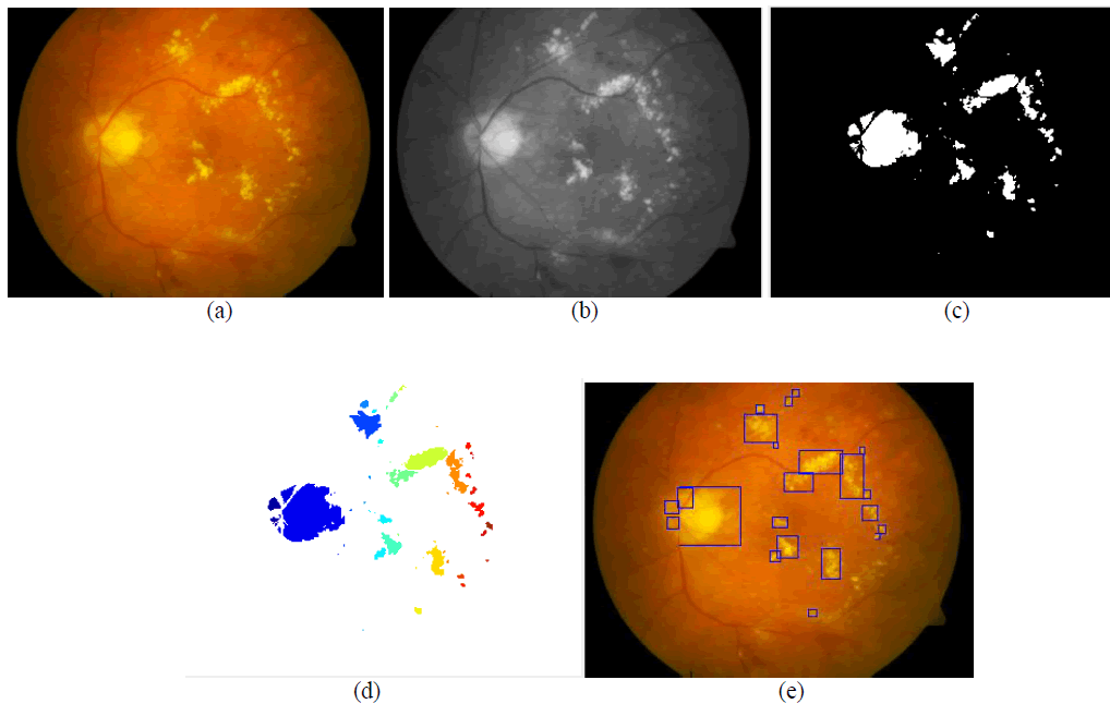 |
| Fig.4.(a) Fundus image (b) Image after intensity conversion (c) Image after thresholding (d) Image after labeling (e) Image after segmentation |
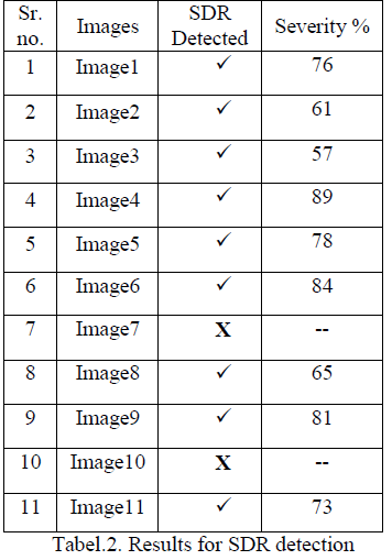
| We tested our algorithm on several images taken from ophthalmologist, and obtained the results for proliferative diabetic retinopathy(PDR) and severe diabetic retinopathy (SDR) as shown below in Table.1 and Table.2 |
 |
 |
CONCLUSION |
| `In this paper,we explore methods towards the development of an automated system for the purpose of detecting Diabetic Retinopathy. This work determines the presence of diabetic retinopathy by applying techniques of digital image processing on fundus images taken by the use of medical image camera by a medical personnel in the hospital.We tested our technique on number of fundus images. This method gives clearer and more accurate output for ophthalmologists,and automated retinal image diagnosis. It also gives severity in percentage.The proposed system demonstrates a high sensitivity and specificity. |
ACKNOWLEDGMENT |
| We express our deep sense of gratitude towards respected Prof. S.U.Bhandari (Head of the E&TC Department) due to her valuable guidance. We are also thankful to Prof.A.M.Fulambarkar for their involvement and their interest in the project.We would like to acknowledge the contribution of our colleagues.We are also thankful to Dr.Santosh S.Bhide,who spent his numerous hours discussing ophthalmic imaging and image analysis with us. |
References |
|