ISSN ONLINE(2319-8753)PRINT(2347-6710)
ISSN ONLINE(2319-8753)PRINT(2347-6710)
Neha, Rani Kumar1, Sanjeev Lalwani2, Manjul Jain3 and RenuDhingra4
|
| Related article at Pubmed, Scholar Google |
Visit for more related articles at International Journal of Innovative Research in Science, Engineering and Technology
The exact knowledge of the topographical anatomy is not only a pre-requisite but also facilitates accurate clinical diagnosis during MRI, CT and ultrasonography. It becomes more fascinating if the anatomical specimen being used to study is dry, odourless, non toxic and not a wet specimen with formalin fumes. Such specimens can be procured by using a novel processing technique called plastination. Thus the present study was undertaken to prepare coronal and sagittal knee region plastinates for studying sectional anatomy of knee joint and to compare them with MRI images of the same. A total of 4 knee joint specimens were collected, washed, cleaned and fixed in 5% formalin. Coronal (1cm) and sagittal (1 cm) slices were made, plastinated using S-10 silicon technique and compared with MRI images of the same. All the structures were exactly corresponding to the MRI images. Thus plastinated coronal and sagittal sections of the knee are ideal as teaching tool for studying sectional anatomy of knee not only because of their instructional value but their durability, ease of handling, transportation to operation theatre and a ready reference material at the work place.Plastinated knee specimens can serve as excellent educational tool for the undergraduate and postgraduate students of anatomy, radiology and orthopedics as they are dry, odorless, nontoxic with good structural preservation and higher instructional value. Fresh knee region when plastinated were esthetically superior in terms of color, dilatation and flexibility thus making them ideal for teaching and hands-on experience.
Keywords |
| Cross-Sectional Anatomy; MRI; Knee; Plastination, S-10 |
INTRODUCTION |
| Sectional anatomy has had a long history, which reached a zenith in the nineteenth century only to decline in the early twentieth, and is now enjoying a renaissance because of advances in radiological techniques such as computed tomography and magnetic resonance imaging. This necessitates the need of including teaching of sectional anatomy as an integral part of medical curriculum at various levels [1]. |
| However formalin fixed wet sections deteriorate with time [2,3]. To overcome these problems we decided to plastinate sections of various anatomical specimens as these specimens offer several advantages over other methods of preservation because they are anatomically precise, clean, dry and easy to handle [4]. Plastinated slices provide an excellent tool for teaching anatomy, pathology and radiology [5-7] for patient education, and potentially as an augmentation to MRI and CT analysis [8,9]. These plastinated sections show anatomical structures in multiple planes and are effective for teaching anatomical spatial relationships, something that students often find difficult to comprehend [10]. |
| Knee joint being the largest, most complicated joint, vulnerable both to acute injury and osteoarthritis, understanding its anatomy is fundamental in understanding any subsequent pathology. A review about image analysis of the anterior cruciate ligament (ACL) anatomy and its application to ACL reconstruction surgery reported that three-dimensional image analysis of the ACL anatomy and its application to the navigation system is becoming more prevalent and reliable for advancing the anatomic studies related to the native ACL and the ACL reconstruction procedure. Thus Plastinated knee sections can not only be of immense help in radiographic analysis but can also provide hands-on experience to various orthopedicians. Thus the present study was undertaken to prepare plastinated coronal and sagittal sections of knee region and compare them with the MRI images of the same which to the best of our knowledge has not been reported in the literature so far. |
MATERIALS AND METHODS |
| Knee specimens were collected from the cadavers being used in the Department of anatomy, AIIMS, New Delhi. Each specimen (25 cm in length) was thoroughly washed with running water and rinsed with normal saline injection through the femoral artery followed by heparin (25,000 I.U. in 5ml) diluted with distilled water in a proportion of 0.2 in 5ml. 5% formalin was injected through the femoral artery for fixation. Specimens were submerged in 5% formalin for 1-2 weeks for a better fixation. Formalin fixed specimens were washed in running tap water for one to two days to remove the excess formalin, cooled to -40°C in order to harden them up and then sliced in coronal (1cm) and sagittal (1cm) planes with the band saw. Cut sections were transferred to 100% cold acetone at -20°C. Acetone concentration was measured daily with acetonometer and acetone changes made when concentration reached around 90%. Dehydration was considered complete when water content was below 1% for three consecutive days. After this specimens were immersed in the silicone resin S10 with S3 Catalyst for 24 hours [11]. They were then subjected to a forced impregnation where the pressure was reduced gradually by using a vacuum pump. The rate of impregnation was monitored by the rate of acetone bubbles which came on the surface. The specimens were kept immersed in the polymer for one day before taking them out. They were finally gas cured in a curing chamber by passing S6 vapour. After the specimens were dry on touch, they were enclosed in polythene bags for the final curing. The various sections demonstrated the internal features and were compared with MRI images of the same. |
RESULTS |
| The work resulted in coronal and sagittal sections of `knee, exactly corresponding to the MRI images serving as teaching material for radiologists without any potential and chemical hazards, especially from formaldehyde. They provided excellent anatomical bone detail, demonstrating organ position, shared structures, and vascular anatomy. The specimens could be stored in the radiology department without wet formalin jars for ready reference. |
[A.] In the coronal slices of knee region the following structures were seen: |
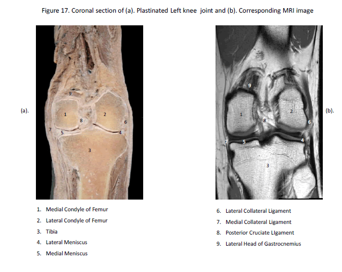
| Poster ior -to-anter ior coronal anatomic sect ions demonst rated the poster ior capsule, the popl i teus tendon, the cruciate l igaments and menisci , the col lateral l igaments and the extensor mechanism [Fig.1]. This plane also displayed the poster ior femoral condy les, which are common si tes of ar t icular erosions. The cruciate l igament s, al though displayed to best advantage in the sagi t tal plane, were also ident i f ied on coronal sect ions [Fig.1]. The obl ique popl i teal l igament and arcuate popl i teal l igament def ined t he poster ior capsule. |
| The LCL ( f ibular col lateral l igament ) was seen as a cord st retching f rom i ts inser t ion on the f ibular head to the lateral epicondyle of the femur . I t was separated f rom the lateral meni scus by the thicknes s of the popl i teus tendon. At the level of the femoral condyles, the meniscofemoral l igament s ( the l igaments of Wr isberg and Humphrey) were observed as thin bands extending f rom the poster ior horn of the lateral meni scus to the lateral sur face of the medial femoral condyle. The PCL was seen as a ci rcular st ructure on anter ior and mid -coronal sect ions. On poster ior coronal images, the t r iangular at tachment of the PCL was di f ferent iated as i t fanned out f rom the lateral aspect of the medial femoral condyle. The MCL, or t ibial col lateral l igament , was ident i f ied on mid -coronal sect ions [Fig 1.] anter ior to sect ions in which the femoral condyles appeared to fuse together wi th the distal metaphysis. The MCL was seen as a band extending f rom i ts femoral epicondylar at tachment to the medial t ibial condyle. I t consisted of super f icial and deep layer s at tached to the per iphery of the medial meni scus. |
| The body and the anter ior and poster ior horns of the medial and lateral menisci were seen as dist inct segments and not as opposing t r iangles as on sagi t tal sect ions. |
[B.] In the sagittal section of knee region the following structures were seen: |
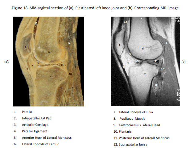
| Bony structures- Patella, Lateral condyle of femur and lateral condyle of tibia [Fig.2] |
| Muscles- Popliteus, Gastrocnemius lateral head and Plantaris [Fig.2] |
| Menisci and ligaments- Patellar ligament and both the horns of lateral meniscus [Fig.2] |
| Infrapatellar fat pad and suprapatellar bursa were nicely seen in the mid- sagittal section [Fig.2]. Sect ions in the sagi t tal plane are key in evaluat ing meni scal anatomy for both degenerat ions and tear s. |
| Other st ructures observed in the sagi t tal plane were the posteromedial and posterolateral corner s, the patel lar and quadr iceps tendons, Hof fa's fat pad and pl icae. |
| The patel lofemoral compar tment , quadr iceps, and patel lar tendon were demonst rated on midsagi t tal sect ion [Fig.2]. The suprapatel lar bur sa (pouch) extended 5 to 7 cm proximal to the super ior pole of the patel la [Fig.2]. Super f icial medial dissect ion displayed the conjoined pes anser inus tendons ( semi tendinosus, graci l i s, and Sar tor ius) . On the lateral aspect of the knee, the LCL and the more poster ior ly located fabel lof ibular l igament ( s t ructures of the posterolateral corner of the knee) we re seen. |
| • The ACL and PCL were best displayed on sagi t tal sl ices. The LCL, or f ibular col lateral l igament , and the biceps femor is tendon were seen on per ipheral sagi t tal sect ions. . |
| • On medial sagi t tal sect ion, semimembranosus tendon and muscle was seen po ster ior ly. The vastus medial is muscle made up the bulk of the musculature anter ior to the medial femoral condyle. The anterolateral femoral ar t icular car t i lage was f requent ly the si te of ear ly erosions or at tenuat ion in osteoar thr i t is ( t rochlear groove ch ondromalacia) . |
| • On medial sl ice approaching the intercondylar notch, the separate anter ior and poster ior horns of the medial meniscus were seen. When sagi t tal sect ions were viewed in the medial to lateral di rect ion, the PCL was seen before the ACL. |
| • Por t ions of both cruciate l igaments were observed on the same sagi t tal sect ion. On midsagi t tal sect ions, the quadr iceps and patel lar tendons were seen. On intercondylar sagi t tal sl ice, the popl i teal vessels were seen in long axis, wi th the ar tery in an anter ior and the vein in a poster ior posi t ion. |
| • On ext reme sagi t tal sect ions, the conjoined inser t ion of the LCL and the biceps femor is tendon on the f ibular head were ident i f ied. The lateral head of the gast rocnemius muscle was seen poster ior to the f ibula. The po pl i teus tendon was seen between the capsule and the per iphery of the lateral meniscus. |
| All the structures were exactly corresponding to the MRI images. The plastinated slices displayed superior differentiation between musculature compared to the scans. Thus, the coronal and sagittal sections of the knee are ideal for studying sectional anatomy of knee which is needed for diagnostic as well as for research and teaching purpose. |
DISCUSSION |
| Anatomy is a fundamental educational science in Medical Universities. It has been involved in radiological instructions for medical students for decades [1]. Most clinicians view internal anatomy with the aid of radiographic images and procedures. Proper interpretation of these images presupposes a detailed knowledge of anatomy. Anatomy teachings should implement a number of clinical procedures, pathological observations and diagnostic methods into the course resulting in a structured program of clinically based teaching of gross anatomy to second- and third-year medical students. The recently developed method of E12 epoxy resin sheet plastination has expanded the learning process twofold, firstly as a developmental microscopy teaching aid enabling us to link histological slides directly to gross anatomical specimens and secondly as a radiographic training tool in the correlation of clinical imaging techniques of magnetic resonance (MRI) and computed tomography (CT) with the teaching of gross anatomy. The direct comparison between E12 serial sectioned cadaver specimens and the equivalent MRI and CT images provides the student with a much clearer understanding of anatomical structures in relation to clinical diagnosis [9]. Radiography has proved particularly valuable in the detection of the early stages of deep-seated disease, when the possibility of cure is greatest. During these early stages there is little departure from the normal. Hence knowledge of the earliest detectable variations, that is, of "the borderlands of the normal and early pathological..." is of great medical importance. Radiographic diagnosis is the most important method of non-destructive testing of the living body. With the rise of modern imaging methods, the demand for sectional anatomy has increased and requires that it should be integrated into medical curriculum as vital component during undergraduate and postgraduate levels. Such sections should also be available in later phases of the curriculum when students deal with patient-oriented material. |
| In the study of anatomy, the use of gross specimens and other biological material is mandatory. Decay of this material is an impediment to all morphological studies, teaching, and research. Thus essential part is an excellent preservation of the biological material being used in teaching for it to be used as an educational tool. Plastinated specimens are clean and odorless, require minimal aftercare & can be stored on shelves or in display cases for a longer duration. As fully cured specimens are durable and do not require 'wet' storage, plastination provides us with the opportunity both to extend the life of this material, and additionally maintain effective student/specimen interaction. The dry plastinated specimens also become transportable. |
| As knee joint is one of the most important joint of human body, understanding the anatomy of the joint is fundamental in understanding any subsequent pathology. The exact knowledge of the topographical anatomy of knee joint is not only a prerequisite but also facilitates the accurate clinical diagnosis of various knee disorders using imaging techniques like MRI, CT and sonography. Interpretation of radiological images need anatomical details in multiple planes, something that students often find difficult to master [12]. In the present study the coronal and sagittal plastinates of the knee joint corresponded well to the MRI images and thus are ideal for studying sectional anatomy of knee for understanding its pathology for clinical diagnosis. In future they will be preferred over wet specimens not only because of their instructional value but their durability, ease of handling, transportation to operation theatre, a ready reference material at the work place and positive impact on learners. |
| Currently we have plastinated the 1cm thick sections by S-10 procedue. We are planning to prepare in future 4-5mm thick sections by epoxy technique which gives more accurate details. |
CONCLUSION |
| Sheet plastination is currently used to produce anatomical sections of different body structures, allowing one to study and teach their topography in an anatomically correct state. Correlation with computed tomography (CT) and magnetic resonance imaging (MRI) techniques gives more insight into their anatomy. This combined approach provides a unique anatomical insight and is a valuable addition to other teaching tools used by medical students, radiologists and anatomists. |
References |
|
 |
 |