ISSN: 2319-9865
ISSN: 2319-9865
1Department of Anatomy, GMERS Medical College, Valsad, Gujarat, India
2Department of Preventive and Social Medicine, SMS Medical College, Jaipur, Rajasthan, India
Received: 01/02/2013 Accepted: 07/02/2013
Visit for more related articles at Research & Reviews: Journal of Medical and Health Sciences
Determining the sex of human skeletal remains, using the skull is important to the disciplines of human osteology, forensic anthropology, paleopathology and paleodemography as compared to other bones in the body namely the human pelvis and long bones. The skull offers a high resistance to adverse environmental conditions over time, resulting in the greater stability of the dimorphic features as compared to other skeletal bone pieces. Moreover, some authors have highlighted the importance of the petrous portion of the temporal bone and its general preservation in cases of extreme unfavourable conditions. The purpose of this study was to determine the existence of sexual dimorphism in the dimensions and the area of mastoid triangle related to the 3 craniometeric points: porion, asterion and mastoidale. A total of 100 skulls, 50 male and 50 female were analyzed. On each skull the 3 craniometeric points were marked on each side. The area (mm2) for each side of the skull right (D) and left (E) was determined and the total value of these measures (T) was calculated. All the lineal dimensions, Porion-Asterion, Asterion-Mastoidale, Mastoidale - Porion, were found significantly higher in males than females with p < 0.01. The analysis of the differences between the sexes in the areas studied for all the 3 areas (Right, Left and Total area) was significant with p < 0.01 Regarding, the total area, which was the preferred measurement because of the asymmetry between the sides of skull, the value of the mean was 1475.69 mm2 for males and the valve of mean was 1288.90 mm2 for females. This study demonstrates a significant result in the 3 studied areas, (D), (E), (T). The total area being the choice as it would avoid asymmetry between the areas and therefore can be used for sexing of human skulls. For the population studied, values of the total area that were greater than or equal to 1599.73 mm2 belonged to a male crania (with 95% confidence). Similarly values for this area less than or equal to 1070.66 mm2 belonged to female crania (with 95% confidence).
Mastoid Process, Forensic Anthropology, Sex determination
Historically, human identification is one of the most challenging subjects that man has confronted. According to Alves [1] identity is a set of physical characteristics, functional or psychic, normal or pathological, that defines an individual.
Human identification is a universal process which is based on scientific principles. Application of the knowledge of physical anthropology for the purpose of forensic medicine constitutes forensic anthropology [2].
The studies for sex determination are based on the dimorphism between the sexes that is present in the majority of human bones.
Most authors emphasize the dimorphism of the pelvis and skull. Krogman with Iscan [3] state that determination of sex, age and race in a collection of 750 skeletons was possible, with levels of reliability of 100% when whole skeleton was present, with 95% reliability when using pelvis alone, 92% using the skull alone and 98% using both the pelvis and the skull.
This clearly demonstrates the importance of these regions – the pelvis and skull for sex determination in forensic anthropological examination.
Bass [4] says that the skull is probably the second best region of skeleton to determine the sex. A great many researchers have studied the dimorphism of the mastoid process between the sexes through the use of its measurements, in isolated form or through the product between its values, emphasizing in a general way that the mastoid process is larger in the male.
In the skull, the temporal bone is highly resistant to physical damage, thus it is commonly found as remainder in the skeletons that are very old, and of this the petrous portion has been described as important for sex determination due to its craniometeric characteristics.
Paiva and Segre [5], introduced an easy technique for determining sex, starting from temporal bone, with a small observational error and a high predictability degree. They found significant differences in the area between the right and left mastoid triangles when comparing male and female skulls, but owing to the asymmetries present in the skull, they recommended observing the value of total area (adding left & right sides) which was also significant, so that when it was higher or equal to 1447. 40 mm2, the skull was diagnosed as a male skull and a value 1260.36 mm2 or less was indicative of female skull.
Ongoing through the available literature, we can recognize the following and hence the importance of using mastoid process for sexing of human skull
a) Importance of skull for sex determination next to pelvis.
b) The importance of temporal bone for anthropological studies due to its robustness and location, making it usually possible to be examined as a fragmented or burned bone.
c) Superior results demonstrated in studies that make use of multiple measurements rather than an isolated measurement of mastoid process to determine sex of the skeleton.
d) Previous studies giving significant results demonstrating the use of mastoid process over other parts of skull in classifying the sexes better.
e) Scarcity of Indian national studies utilizing the aforementioned work of great authors and forensic anthropologists.
Thus, the present study, which was carried out using resources generally available to the majority of medical examiners, forensic experts and physical anthropologists, is founded on an easily applied methodology with the purpose to determine the existence of sexual dimorphism in the dimensions and the area of mastoid triangle, measured directly on the skull.
This study was conducted on 100 adult skulls of known sex (50 male and 50 female) collected after excluding those skulls that presented evidence of traumaor deformations from the departments of anatomy at Indira Gandhi government medical college Nagpur, Geetanjali medical college, Darshan dental college and Pacific dental college, Udaipur. On each skull, the 3 craniometeric points were located and marked by a single investigator (who was blind about sex of skulls) on both sides of the skull.
A) Porion (PO) : Superior most point of the external acoustic meatus.
B) Mastoidale (MA) : Inferior most point of the mastoid process.
C) Asterion (AS) : The meeting point of lambdoid, occipitomastoid and parietomastoid sutures. (Fig.1)
Linear measures were carried out directly using a sliding vernier caliper (with a least count of 0.01mm). The mastoid triangle area was calculated by means of the Herons’ formula.
The values in mm2 in the present study were obtained by calculating the area of the demarcated triangle on each side of the skull, viz., right area (D) and left area (E). The total area (T) is the sum-total of these two measurements. The results were subjected to statistical analysis and were interpreted subsequently.
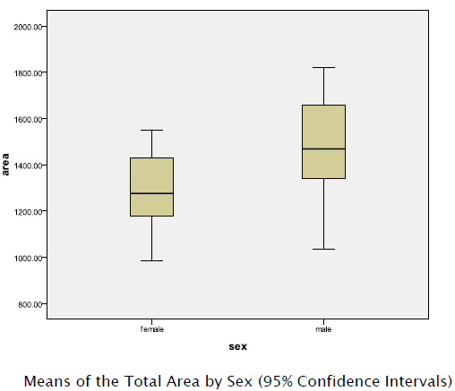
In all the 100 analyzed skulls, all of the lineal dimensions and the calculated areas where higher in males than in females. This data when put to statistical analysis, proved the significant difference with p < 0.01 in all the values calculated in males when compared with that of females.
The maximum values for the total area calculated in the males and females were 1820 mm2 and 1549 mm2, respectively, whereas the minimum values for the same were 1035 and 986.7 mm2 in males and females, respectively.
The values for the means of lineal dimensions and the calculated areas (right, left and total areas) observed in males and females are presented in Table 1.

The analysis of the mastoid process characteristics is important in the determination of sex for forensic purposes. Many authors agree that qualitative aspects, such as their size, ruggedness for muscular attachment or mastoid process inclinations are very good indicators of sexual dimorphism.
The objective of this study was to demonstrate that through an easily applied methodology, majority of the forensic anthropologists, coroners and medical officers at primary centers would be able to determine the sex of various skulls.
This study analyzes the dimensions of the denominated mastoid triangle, defined according to that described by De Paiva & Segre, but measured directly on the skull.
Our values for the male mean total area was less as compared to that of De Paiva and Segre, whereas for the female these values were greater. It is necessary to keep in mind that in the original method of De Paiva and Segre, one obtains the lineal dimensions based on a plane image by means of a xerographic copy of a structure convexity (skulls), which diminishes the distance between the points; hence some variations are seen when compared with these values.
Since this study was based on anthropometric techniques, it surpasses in importance the older studies such as those of Broca [6] and Martin [7]. It also improves on the criteria reported by Bass, which were based only on descriptive anatomical aspects.
Here in the present study, by using a measurement of the surface area, or in other words, by using the result product between 2 values, our results improve upon those studies by Schultz [8], Schaefer [9] which used only single measurements.
The mastoid region used in this study, being a part of temporal bone, is recognized as being the most protected and resistant to damage, due to its anatomical position at the base of skull, these findings have been reconfirmed by many authors Kloiber, Weels, Gejval, Spence as cited by Wall and Henke [10].
Therefore, compared with the most important historical studies dealing with the sex determinations of skulls, the present study shows improved results. These results are based on anthropometric techniques, and open paths for further studies based on statistics, which could be of considerable aid to medico-legal investigation.
The required equipment for the execution of this technique is readily available to the majority of forensic experts, medical officers. This technique is easy to execute, offers quick results, and dispenses with any type of special training for medical examiner.
The technique for sexing skulls presented in this study offers a practical alternative to other methods and meets the needs and realities of the forensic investigation in our country today.