ISSN ONLINE(2319-8753)PRINT(2347-6710)
ISSN ONLINE(2319-8753)PRINT(2347-6710)
Chavitra.V 1, Suganya.S 1, Susi.S 1, Uma Maheshwari.K 1, Mr.Solaiappan solasubbu M.S 2
|
| Related article at Pubmed, Scholar Google |
Visit for more related articles at International Journal of Innovative Research in Science, Engineering and Technology
Embedded systems are growing vast in medical devices. In this paper, we segment tooth images based on feature contour algorithm in which the images are captured through dental intraoral USB camera. The dental intraoral system supports the capturing of dental images and image tooth segmentation. This device helps dentists in diagnosis with the help of dental images captured by dental camera. In this system, we propose advanced characteristics for the dental intraoral system including touch screen with graphic user interface (GUI), dental image processing, patient records, and dentist’s diagnosis note. Especially, the segmentation of tooth is important for examining and extracting tooth characteristics from segmented images. On Feature contour without edge algorithm has been proposed in this paper for the segmentation of the tooth. Moreover, our system is portable, economic and ready to be applied at dental clinics. It also helps dentists examine at patient’s home and voyages, apart from clinics.
Keywords |
| Dental intraoral system, dental Image, tooth segmentation, ARM, Micro2440, embedded system, Linux, opencv. |
INTRODUCTION |
| Embedded systems with ARM microprocessor have become widely use in medical systems due to low power, less time consumption and high performance. Many medical applications are based on image acquisition systems [8], [6]. Such kinds of systems are used in X-ray scanner, computed tomography (CT) scanner, magnetic resonance imaging (MRI), and dental intraoral system. The image acquisition system mainly supports three functions, i.e. image capturing, processing and displaying results. For example, Yong-lin LIU proposed system shows an image acquisition processing system using ARM and Linux [5]. In this system, the embedded image processing system consist the functions of image capturing, displaying and processing. In this design, Yuanpig Ni proposed an image acquirement with the technique of Bluetooth [7]. This design gains images from USB camera and transmits those images to medical data centre. Dental intraoral design is one of image acquisition applications. KAB dental [3], Miharu [2] have presented systems which can gain tooth images and display them on LCD. However, the current systems do not support touch screen GUI, dental image processing and patient record. Our system is an embedded design for dental image processing. In this system, ARM microprocessor running on embedded Linux. Our system is supported by the libraries of open-CV. This system has following functions: |
| Capturing dental image Touch screen GUI Dental image processing including brightness, contrast and tooth segmentation Patient records and dentist’s diagnosis note |
II. SYSTEM DESCRIPTION |
| The system is divided into 2 parts: hardware and software. Hardware is built based on Friendly ARM Mirco2440 board. Dental images are captured from a USB dental camera and displayed on LCD touch screen. An embedded program is developed by using opencv. The program supports functions such as, dental image processing, patient’s data storage and supports dentists for diagnosis by dental images. Software is built in Embedded Linux environment. Dental images are processed by using Linux. ARM microprocessor is used which runs on embedded Linux Operating system. |
III. HARDWARE DESCRIPTION |
| The hardware is based on Micro2440 and it is a ARM9 development board and has many applications in embedded systems. Micro2440 uses S3C2440 microprocessor which is a 32-bit microcontroller fully integrated as a core controller.S3C2440 has interface with, USB Camera (via USB port), PC (via RS232 port) and Ethernet. Besides, touch screen LCD, camera and memory are equipped. Camera unit capture images and send them to S3C2440 via USB port. The 1.3M Dental Intra-Oral USB Camera which is small, high sensitivity and high definition has been used in our system. Dental images are sent to CPU and are showed on LCD unit. |
 |
IV. SOFTWARE DESCRIPTION |
| The software system consists of 4 parts, namely Bootloader, Embedded Linux, Library and Application. Bootloader is started when the power is on. Bootloader helps hardware to transport the kernel of the operating system into RAM memory. The Linux kernel and Root File System is loaded into system memory. Linux kernel is a core of operating system (OS) which contains controlling functions of hardware. The Kernel Linux-2.6.32.2 which is suitable for Mirco2440 board is used in our system. Open CV (open computer vision) is an open source library and it plays an important role in research and development of computer vision area. It serves for many fields such as security image processing, medical image processing, surveillance camera, identification, and robot programming. The embedded system is divided into two main parts: Graphic User Interface and Dental Image Capturing and Processing. Graphical user interface helps dentists to manage patients easier. Graphical User Interface includes functions: patient information, diagnosis note box, data and time management, Virtual keyboard, and data storage. The dental image capturing and processing consists of brightness, contrast, and tooth segmentation. The dental images are acquired from a dental camera through USB interface, and then stored in by using opencv library. |
V. TOOTH SEGMENTATION |
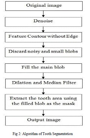
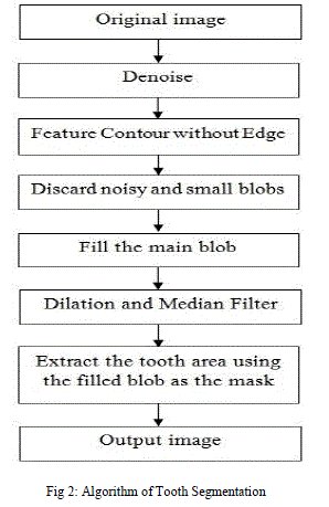
| The input RGB image to be segmented is taken and is converted to grayscale image. This makes the image effective for further process. Gaussian noise generally appears in the medical image. Weiner filter is used to remove Gaussian noise. The tooth features in an image is employed to segment by using feature contour extraction [1]. To find the full mask of the tooth, some additional computational steps including discard noisy and small blobs, fill blobs need to be applied to the global region image. Dilation is a convolution of some image (or region of an image).Dilation operation and median filter are applied to the image region to expand the mask and smooth the contour of the mask. Dilation expands region & the exact result will depend on the kernel. The median filter replaces each pixel by the median or “middle” pixel value in a square neighborhood around the center pixel. Median filtering is able to ignore the outliers by selecting the middle points. After tooth segmentation process, the region outside the tooth area is removed. |
 |
VI. IMPLEMENTATION AND RESULT |
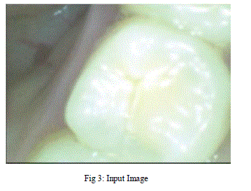
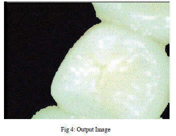
| The dental intraoral system implemented on Micro2440 board using Embedded Linux has been presented in this paper. The diagnosis of tooth has been carried out on PC and Micro2440 board for the storage of data, additional storage facility can be included. Here, we are using RAM memory for storage of patient information. Compared to other system our system is portable, it can be carried away anywhere. It is easy to use in dental clinics. The tooth image captured from the camera and the segmented image are shown in the figure 3 and figure 4, respectively. Consequently, the proposed system is very useful when being applied to dental clinics. |
 |
 |
VII. CONCLUSION |
| Dental intraoral segmentation is based on Micro2440 and Embedded Linux was described. The function of our system includes dental image capturing, tooth image segmentation, management of patient information and storage of data. The dentists and patients can observe dental images in real time LCD touch screen. The system is portable and helps dentist to diagnose at patient home and voyages. |
References |
|