ISSN: 2319-9865
ISSN: 2319-9865
Visit for more related articles at Research & Reviews: Journal of Medical and Health Sciences
Variation of musculocutanous nerve at the level of brachial plexus is common. The musculocutaneous nerve is a branch from lateral cord of brachial plexus. It supplies flexor muscles of the elbow and skin on the lateral aspect of forearm. During routine dissection of the upper limb of a seventy year old adult male cadaver, a communication between medial and lateral cord of brachial plexus in the left axilla was noted. On the same side in the arm, musculocutaneous nerve was absent. The muscles supplied by musculocutaneous nerve were innervated by branches of median nerve and a muscular branch to the brachialis muscle continued as lateral cutaneous nerve of forearm. There was no variation in the musculocutaneous nerve on the right side but there were two communicating branches between musculocutaneous and median nerve. Knowledge of such variation has clinical significance in median nerve injury and also helpful in diagnostic clinical neurophysiology, surgical procedures and management of shoulder and arm trauma. Though such variations are common, it is of clinical significance for radiologists, anaesthetists and surgeons besides of being academic interest for the anatomists.
Keywords |
| Brachial plexus, Medial cord, Lateral cord, Neural communication, Musculocutaneous nerve, Median nerve. |
Introduction |
| Anatomical variations involving the branches of brachial plexus are common and have been reported in the literature by many authors. The prevalence of variation ranges from 12.8-53% [1]. But the variation pertaining to roots and cords are uncommon [2].The median nerve, musculocutaneous nerve and ulnar nerve arises from brachial plexus and runs through the anterior compartment without receiving any branch from the neighbouring nerves [3]. Among the communication between the nerves in the arm, the communication between musculocutaneous and median nerve is common [4]. Brachial plexus is the plexi form arrangement of anterior primary rami of lowers 4 cervical and first thoracic nerves in the axilla. It consists of supra clavicular and infra clavicular parts. The supraclavicular part has roots and trunks whereas the infra clavicular part has cords situated in relation to second part of axillary artery namely medial, lateral and posterior cords of brachial plexus and branches from the cords[2].The musculocutaneous nerve (C5,C6,C7) is a branch from the lateral cord of brachial plexus in the axilla. It supplies the muscles of anterior compartment of arm. It leaves the axilla by piercing coracobrachialis muscle and enters the front of arm where it runs between biceps and brachialis muscle. Above the elbow, it pierces deep fascia and continues as lateral cutaneous nerve of forearm [5]. The lateral cord after giving off musculocutaneous nerve continues as lateral root of median nerve which joins with medial root from medial cord of brachial plexus to form trunk of median nerve. Median nerve does not give off any muscular branches in the arm [1].The present case report describes a communication between medial and lateral cord of brachial plexus with absence of musculocutaneous nerve on the left side and muscles of anterior compartment of arm supplied by median nerve and also communications between musculocutaneous and median nerve on the right side. Knowledge of this variation and communicating branches between the nerves is of importance during surgical interventions in the axilla or arm and in diagnostic neurophysiology. |
MATERIALS AND METHODS |
Case Report |
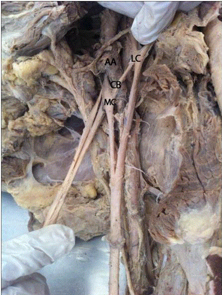
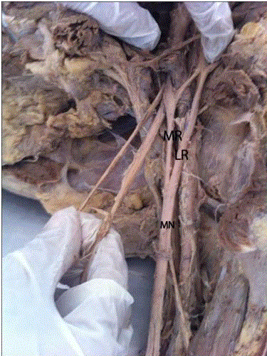
| During routine dissection of the upper limb of a 70 year old adult male cadaver at Sri Muthukumaran Medical College & Research Institute, Chennai, India, following variations in the brachial plexus and its branches were noted bilaterally. In the left axilla, there was a communication between medial and lateral cord of brachial plexus. This intercordal communication ran obliquely over the axillary artery and was about 2.6 cm in length (Fig.1). On the right side axilla no such communication was observed. The lateral cord of brachial plexus in the left axilla did not give off musculocutaneous nerve and it continued as lateral root of median nerve (Fig.2). The coracobrachialis, biceps and brachialis were innervated by branches from the median nerve. |
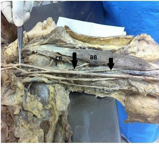
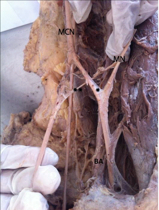
| The muscular branch to biceps arose close to the distal end of coracobrachialis ran laterally and supplied the muscle. Another muscular branch ran between biceps and brachialis supplying the muscles and continued in the forearm as lateral cutaneous nerve of forearm (Fig.3). In the right side axilla, musculocutaneous nerve was seen to arise from lateral cord and there were two communications between musculocutaneous and median nerve. The musculocutaneous nerve after piercing coracobrachialis gave a large first communicating branch to median nerve which was 13.5 cm from the tip of coracoid process ran over the brachial artery and joined the median nerve at a distance of 17.5 cm from the same point. Another small second communicating branch arose from median nerve and ran over the brachial artery and behind the first communicating branch and joined the musculocutaneous nerve distal to first communicating branch (Fig. 4). The remaining trunk of musculocutaneous nerve continued as lateral cutaneous nerve of forearm. Median nerve did not supply any muscles in the right arm. |
DISCUSSION |
| Variations in the formation of brachial plexus have been reported by many authors with varying frequencies of 6.2%, 5.8%, 0.57% and 3%.Renu et al described a case of intercordal communication between medial and lateral cord of brachial plexus which was about 2.2 cm in length with variant branching pattern of posterior cord[2]. Song et al observed a communication between medial and lateral cords on 25 sides among 100 cadavers and mean length was about 1.67 cm[6]. In our study, intercordal communication between medial and lateral cord was about 2.6 cm in length. The anatomical data regarding these communicating branches will be helpful for surgeons who are performing surgical procedures in the neck and axillary region. |
| Le Minor (1992) classified communication between median nerve and musculocutaneous nerve into five types |
| Type I - No communication between median and musculocutaneous nerve |
| Type II - Fibres of medial root of median nerve pass through musculocutaneous nerve and join the median nerve in the middle of the arm |
| Type III- Lateral root of median nerve pass through musculocutaneous nerve and after some distance leave it to form root of median nerve |
| Type IV- Musculocutaneous nerve fibres join the lateral root of median nerve and after some distance musculocutaneous nerve arises from median nerve |
| Type V- musculocutaneous nerve absent and entire fibres ofmusculocutaneous nerve pass out through lateral root and muscles supplied by musculocutaneous nerve branch out from median nerve [7]. |
| In our study, left side variation of absence of musculocutaneous nerve belongs to type V variation since the muscles supplied by musculocutaneous nerve were innervated by median nerve. The complete absence of musculocutaneous nerve and takeover of innervations of muscles in the anterior compartment in the arm by median nerve are uncommon [8]. Absence of Musculocutaneous nerve will not cause paralysis of flexor muscles of the arm and loss of sensation on the lateral aspect of the forearm because the branches are given from other nerves like median nerve, lateral root of median nerve or lateral cord of brachial plexus [9].In our study, there is absence of musculocutaneous nerve and the muscles and cutaneous area supplied by MCN were innervated by median nerve. This variation should be taken into consideration when patients presents with unexpected weakness of elbow flexors and loss of sensation along lateral side of forearm in median nerve injury [10] andduring anaesthetic blocks & surgical approaches in the region [11]. |
| Venieratos and Anangnostopoulou described three types of communication in relation to coracobrachialis muscle. |
| Type I- communication between musculocutaneous and median nerve proximal to the entrance of coracobrachialis muscle. |
| Type II- Communication distal to muscle. |
| Type III- Both musculocutaneous nerve and communicating branch does not pierce the muscle [12]. |
| Choi et al also classified communication into three patterns. Musculocutaneous nerve and median nerve fused in pattern 1, one communicating branch between the nerves in pattern 2 and two communicating branches between the nerves in pattern 3[13]. |
| In our study, right side arm showed two communications between musculocutaneous and median nerve distal to coracobrachialis muscle hence belonging to type II of Venieratos classification and pattern 3 of Choi classification. |
BA- Brachial artery |
| The first communicating branch from musculocutaneous nerve to median nerve has the same diameter as that of lateral root of median nerve hence it may be considered this as third root of median nerve or second lateral root of median nerve. Both the communicating branches ran in front of brachial artery and hence more prone for injury in surgical operations [14]. Communication between median and musculocutaneous nerve is clinically significant when dealing with patients of nerve entrapment syndrome of upper limb and in traumatology of shoulder joint and alsoin diagnostic neurophysiology [15]. It is possible that variation in the current study is the result of developmental anomaly. In humans, forelimb muscles develop from mesenchyme of paraxial mesoderm in the fifth week of intrauterine life. The motor axons arrive at the base of limb bud, they mix to form brachial plexus and the growth cones of axons continue in the limb bud [16].The developing axons is regulated by expression of chemo attractants and chemo repulsants which takes place in a highly coordinated manner. Any alteration in the signalling between mesenchymal cells and neuronal growth can lead to significant variations [17]. This variation may also be due to circulatory factors at the time of fusion of the brachial plexus cords [18]. |
Conclusion |
| Knowledge of these anatomical variations is important for surgeons, clinicians, anaesthetists, radiologists and orthopaedicians to avoid unexpected complications. |
Figures at a glance |
 |  |  |  |
|||
| Figure 1 | Figure 2 | Figure 3 | Figure 4 |