ISSN ONLINE(2319-8753)PRINT(2347-6710)
ISSN ONLINE(2319-8753)PRINT(2347-6710)
T. Mbiadoun Lionel1, Kom Martin2, Ntsama Eloundou Pascal3
|
| Related article at Pubmed, Scholar Google |
Visit for more related articles at International Journal of Innovative Research in Science, Engineering and Technology
In this work we present a universal module for acquisition and transmission of electrophysiological signal. The goal of our work is to design a low cost module which can help in country under development. The developed module is based on a variable gain amplifier and active noise cancellation both controlled by microcontroller. The system is able to acquire various signal, store and transmit to a distant server. Data sent to the server are processed. In the case of heart signal, a result concerning heart rate, heart condition is sent back to the module. Finally the server will automatically sent a medication and inform the physician
Keywords |
| Phonocardiography, ECG, PCG, GSM transmission, telemedicine. |
I. INTRODUCTION |
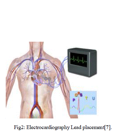
| Hearth diseases in Africa are with HIV and malaria the first cause of death. In those countries as in all poor country or country under development, the number of heart surgeons is very small and they are most of time concentrated in high density urban areas. Heart disease treatment in this case can mean a long and costly transportation to an urban center. This process takes up time and money, both of which are not in large supply for many rural dwellers. A device that can be used in the field to detect heart disease would benefit this specific group of people. By detecting heart disease remotely, we can prevent unnecessary trips, save time and money. In order to develop an understanding of the capabilities and potential scope of this project, a basic understanding of the heart and its mechanics was necessary. As doctors perform auscultations of a patient’s heart, there are some key features that are used in diagnosing cardiac issues. Generally during auscultation, the “lub-dub” valve sounds and blood flow within the heart are the main areas of focus during initial examinations. Electrocardiogram (ECG) may be used by physicians in order to measure electrical current, and therefore the functioning of the heart. The ECG uses multiple leads attached to the patient’s body in order to capture the electrical signals from the heart. This first way or process can be painful for the patient and very expensive too. The use of phonocardiography is a new approach and will be suitable for Africa because they are non-invasive,low-cost and accurate for diagnosing some heart diseases.The advancement of technology has paved the way for signal processing methods to beimplemented and applied in many simple tools useful in everyday life. This is most notablein the medical technology field where contributions involving the intelligent applicationshave boosted the quality of diagnosis [1].The module presented in this paper is able to work with the majority of physiological signal: ECG, PCG, EEG, EGG, EMG, ERG… The main part which is amplification, processing and transmission will remain the same while the only movable part is the input sensor. This work, we will first present some electrophysiological signal, we will present the material used in this work, present our methods and finally give a conclusion. |
II. LITERATURE SURVEY |
| a. ELECTROCARDIOGRAPHY |
| Electrocardiography is the recording of the electrical activity of the heart. Traditionally this is in the form of a transthoracic interpretation of the electrical activity of the heart over a period of time, as detected by electrodes attached to the surface of the skin and recorded or displayed by a device external to the body.[2] The recording produced by this noninvasive procedure is termed an electrocardiogram (also ECG or EKG). It is possible to record ECGs invasively using an implantable loop recorder.An ECG is used to measure the heart’s electrical conduction system. It picks up electrical impulses generated by the polarization and depolarization of cardiac tissue and translates into a waveform. The waveform is then used to measure the rate and regularity of heartbeats. A contact ECG signal (from skin) usually have a voltage amplitude from 0.5 to 4mV and a frequency from 0.01 to 250Hz [3] |
| b. ELECTROMYOGRAPHY |
| Electromyography (EMG) is a technique for evaluating and recording the electrical activity produced by skeletal muscles.[4] EMG is performed using an instrument called an electromyograph, to produce a record called an electromyogram. An electromyograph detects the electrical potential generated by muscle when these cells are electrically or neurologically activated. There are two kinds of EMG in widespread use: surface EMG and intramuscular (needle and fine-wire) EMG. To perform intramuscular EMG, a needle electrode or a needle containing two fine-wire electrodes is inserted through the skin into the muscle tissue. Generally needle EMG have a voltage amplitude from 0.1 mV to 5mV and a frequency of from 0 to 10.000Hz[3]. |
| c. ELECTROENCEPHALOGRAPHY |
| Electroencephalography (EEG) is the recording of electrical activity along the scalp. EEG measures voltage fluctuations resulting from ionic current flows within the neurons of the brain.[5] In clinical contexts, EEG refers to the recording of the brain's spontaneous electrical activity over a short period of time, usually 20–40 minutes, as recorded from multiple electrodes placed on the scalp. EEG is most often used to diagnose epilepsy, which causes obvious abnormalities in EEG readings.[6] It is also used to diagnose sleep disorders, coma, encephalopathies, and brain death. Normal EEG signal has a voltage amplitude from 5 to 200 μV and a frequency from 0 to 150Hz[3]. |
| d. PHONOCARDIOGRAPHY |
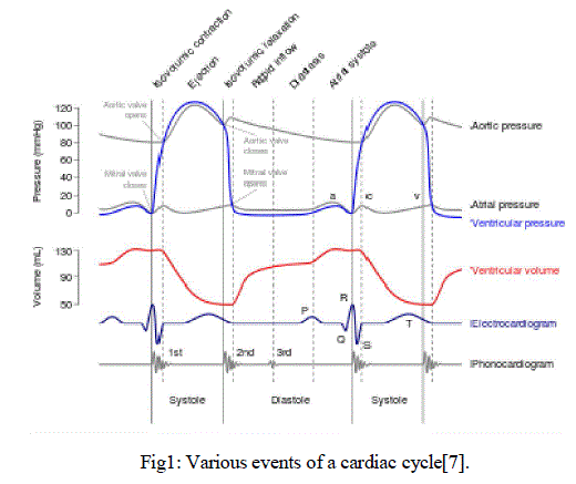
| Phonocardiography is the study of heart sounds. The study of heart sound dates to history. Most of the stethoscopes were acoustic in nature with very less sound amplification. With advent of technology the transition took place into electronic and the more powerful digital-electronic stethoscopes [7].A Phonocardiogram or PCG is a plot of high fidelity recording of the sounds and murmurs made by the heart during a cardiac cycle. The sounds are thought to result from vibrations created by closure of the heart valves. There are at least two: the first when the atrioventricular valves close at the beginning of systole and the second when the aortic valve and pulmonary valve close at the end of systole. It allows the detection of sub audible sounds and murmurs, and makes a permanent record of these events. In contrast, the ordinary stethoscope cannot detect such sounds or murmurs, and provides no record of their occurrence. That is why more sophisticated electronics stethoscope are in study nowadays to always improve the amplification factor necessary for a deep study of heart sound. |
 |
III. MATERIAL AND METHOD |
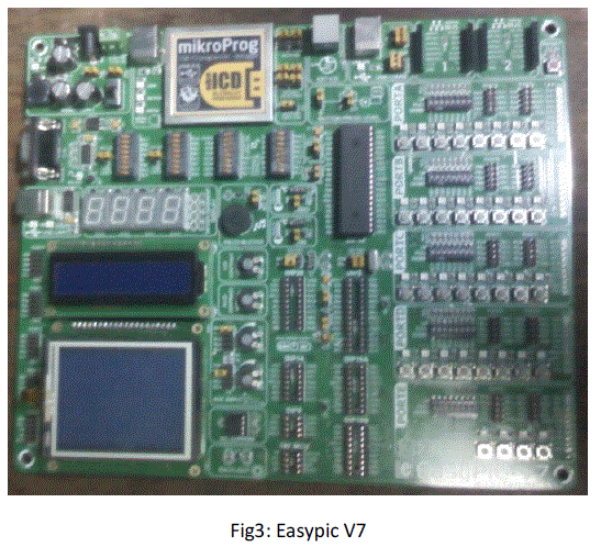
| In this study we used hardware composed of Easypic V7, GSM Click, MP3 Click, microSD Click. The Sofware use to study the result is matlab 7.5. Finally we have developed a server application which receive and analyse the signal from our data base. |
| a. EASY PIC V7 |
| Easypic V7 is a very intuitive development board from mikroElectronica. With the four different connectors for each port, it is easy to connect accessory boards, sensors and other very easy for development. Powerful on-board mikroProg programmer and In-Circuit debugger can program and debug over 250 microcontrollers from microchip manufacturer. It is among few development boards which support both 3.3V and 5V microcontrollers. With the propriatory mikroBUS we can easily connect the mikroClicks (GSM Click, microSD Click…) and develop faster than ever. We have use the microcontrollers 18F4550 which has 16MIPS operation, 64K bytes of linear program memory, 3896 bytes of linear data memory, several timers, ADC converters, comparators and support voltages from 1.8V to 5V.[8] |
 |
| b. GSM CLICK |
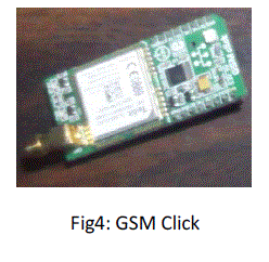
| The GSM click is our transmission and reception module based on Telit GL865-QUAD. The board works with 3.3V and 5V through mikroBUS. The GL865-QUAD modem support GSM/GPRS 850/900/1800/1900Mhz quad band frequency. Additional features such as integrated TCP/IP protocol stack (including UDP, SMTP, ICMP and FTP), serial multiplexer, remote AT commands and many more, extend the functionality of the board [8]. |
 |
| c. MP3 CLICK |
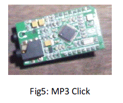
| The MP3Click is an accessory in mikroBus form factor. It includes a stereo MP3 decoder chip VS1053 which can decode multiple fomats (Ogg vorbis, MP3, MP1, MP2, MPEG4, WMA, FLAC, WAW, MIDI), and encode three different formats from microphone in mono or stereo format (Ogg vorbis, IMA ADPCM, 16-bit PCM). The board uses industry standard serial peripheral interface (SPI) for communication, and has built-in stereo headphone and microphone audio connectors. This module was used to acquire and play the heart sound in our system [8]. |
 |
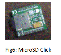
| The microSD click features a micoSD card slot for microSD cards used as a mass storage . SPI interface ensures simple communication at high data rates. It can be used for reading or storing data like music, text files, videos and more. The board is designed to use 3.3v. Our project includes this board to store heart sound and additional data on patients for further use. |
 |
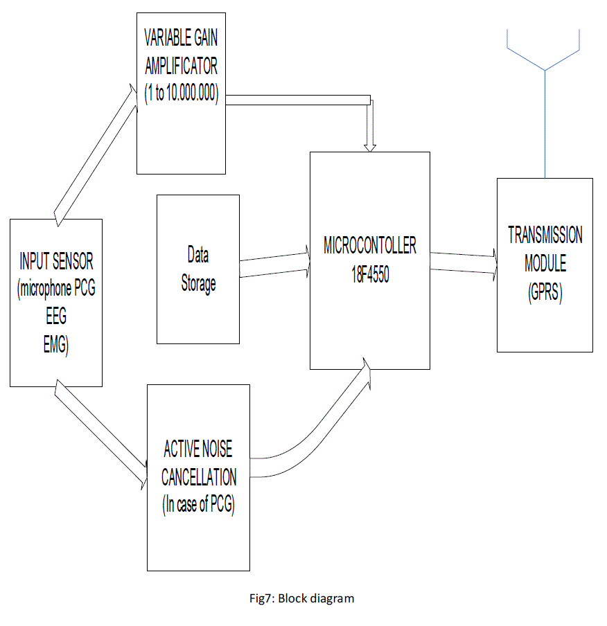
| The work presented in this paper is based on the following block diagram. The input sensor is the movable part and is replace according to the type of signal acquired. For our experimental work we focus on Phonocardiogram which is a representation of heart sound. Our input was based on an electret microphone. To ensure a better acquisition of the signal we have implemented the Active noise cancellation to reduce environmental noise[9][10]. The variable gain amplifier is controlled by the microcontroller according to the input signal received from the sensor. After processing by the microcontrollers, the data is stored in the SD card and real time sent to a distant server using GPRS technology. The sever receives the information, stored it in the database analyse the signal in our case the PCG and send back the result and a medication to the patient in case of pathology. |
 |
| f. SYSTEM FLOW CHART |
 |
IV. RESULTS AND DISCUSSIONS |
| The module based on Easypic V7 has been used during our experimental work. The system acquire, store and send signal to a distant server. The server analyse the signal process it in the case of heart signal, the server is able to give the heart rate and diagnose simple sickness like arrhythmia. |
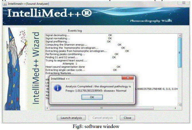
| The following figure shows a windows of our application during a heart sound analysis. This application developed in java is able to analyse heart sound sent by our equipment and give and answer to the patient according to the signal received. Our application name intellimed++ uses neural network to perform the treatment [11]. |
 |
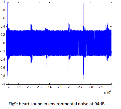
| Figure 9 and 10 show respectively the acquire heart sound with an environmental noise of 94dB and the filtered heart sound. The signal of figure 10 is saved on the circuit in an SD card and is also sent to the distant server for further analysis though GPRS using the telit modem GL865-QUAD. The heart sound is also played and shown on the screen in real time. The response time of our system is 3.2s if there is no network congestion. |
 |
V. CONCLUSION |
| In this paper we have developed a prototype of universal device for acquisition and transmission of electrophysiological signal. We have designed the circuit using Easypic v7 and the main part of this circuit is the microcontroller 18F4550. Our experimental study was focus on phonocardiogram. We have successfully acquire, store and send a sound heart using the circuit and we have successfully apply the active noise cancellation to reduce environmental noise from heart sound. To make sure that the system is working with other electrophysiological signal, we have used a Low frequency generator to test the circuit and we had very good results. As perspective we intend in a future work to reduce the processing time to be closer to the real time processing. |
References |
|