
Pathology 2018
Research & Reviews: Journal of Medical and Health Sciences
ISSN: 2319-9865
Page 60
October 08-09, 2018
Edinburgh, Scotland
17
th
International Conference on
Pathology & Cancer
Epidemiology
I
n case of rhinitis the diagnosis requires a precise diagnostic
procedure and is essentially based on the exclusion of allergies,
infections or defects of the nose. The clinical diffusion of nasal
cytology has allowed recognizing the non-allergic rhinitis. Nasal
Cytology allows identifying non-allergic cell-mediated rhinitis
from neutrophilis (NARNE), eosinophils (NARES) and mast cells
(NARMA). The enrichment technique in the liquid phase also
allows improving the quality of the sample making it easier to
read by the pathologist. The poster describes the technique used
to set up the samples and the results obtained.
Biography
Gozzi Michela, born in 1973, obtained a degree in 1997 from the Faculty
of Medicine and Surgery of Brescia in a Biomedical laboratory techniques
with a score of 110/110 with honors. From 1997 to 2005 she worked at the
Service of Pathological Anatomy of the Poliambulanza Foundation, then she
worked as co-ordinator of the Cytology service of the OxigenLab medical
analysis laboratory. From2014 she startedworking as a freelance cytologist
in the cytopathology service of the Clinical Institute of the City of Brescia.
biagio.biagio_mg@libero.itUse of the cytological method of liquid phase in nasal cytology
Gozzi Michela, Sergio Fiaccavento
and
Cordoni Rosangela
Istituto Clinico Città Di Brescia, Italy
Gozzi Michela et al., RRJMHS 2018
Volume: 7
17
th
International Conference on Pathology &Cancer Epidemiology
October 08-09,2018 Edinburgh, Scotland
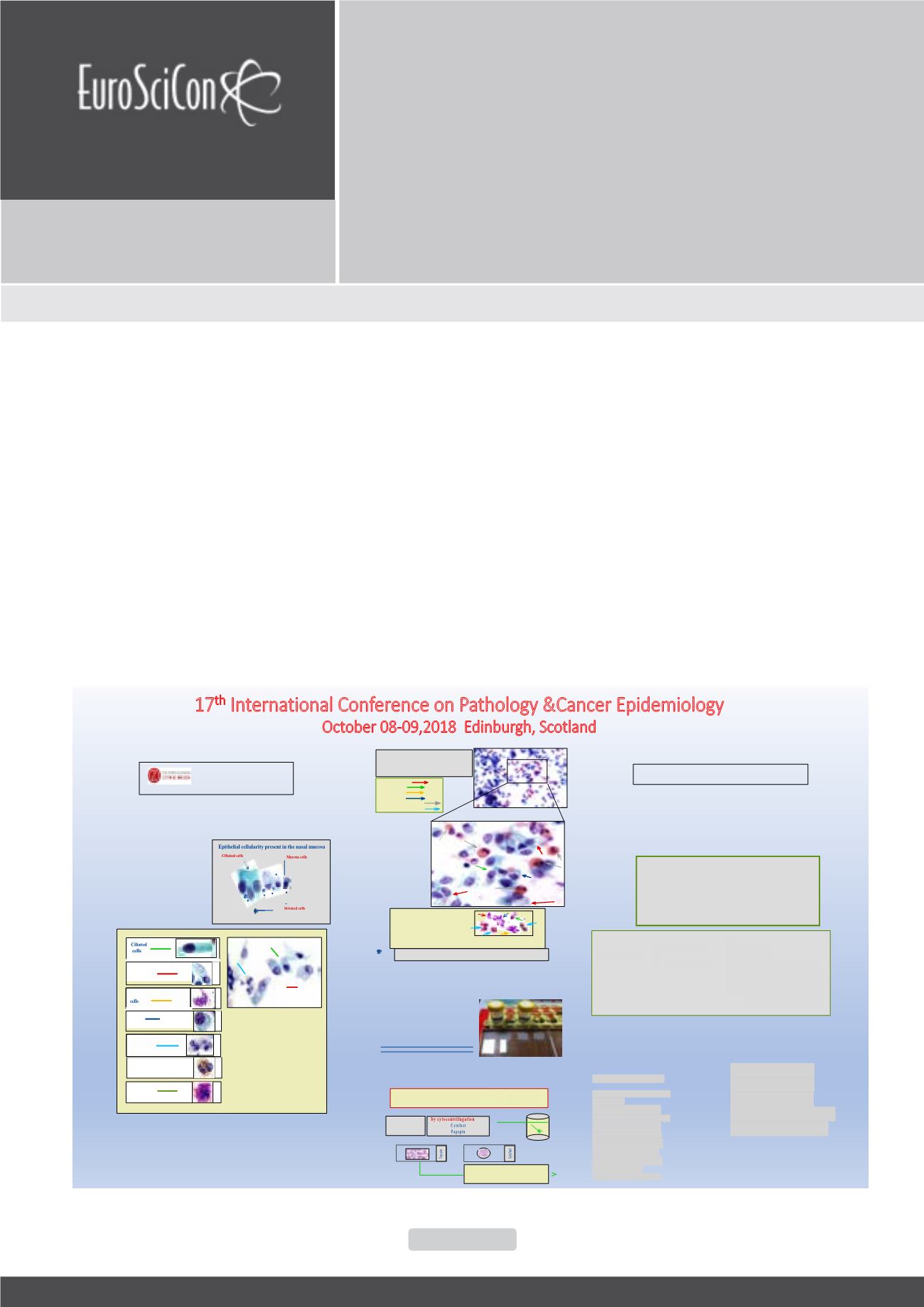
RHINOCITOGRAM:
l
Ciliates cylindrical cells : ++
l
Muciparous globet cells :+++
l
Striated cells:++
l
Basal cells: ++
l
Neutrophilsgranulocytes :+
l
Eosinophils granulocytes :++
l
Mastocytes: 0
Comment
:
in the absence of significant inflammation and in the presence of numerous eosinophils,
the increase of muciparousglobet cells, is such as to suggesta picture of mucin
ous
metaplasia
.
Nasal cytology : C 770/18
Clinical news:
Pale rhinopathy (vasomotor component) with upper
left cicatricial deflection and tendency to substenosis
of themeatal area of this side, in a woman aged 44,
previously operated of
septoplastic
and with nasal
pyramiddeviate fromprevious trauma.
Muciparous cells
Ciliated cells
Basal cells
Striated cells
Granulocytes eosinophil
Granulocytesneutrophil
MGG
The samplingmethods used by various author are different.What
we use is simple and consists of scraping carried out at the level
of the nasalmucosa, bilaterally, at the inferiore turbinate.
Material used
:
In a first sampling with cytobrush, 3 glasses
with traditional smears are prepared for
coloring according toMGG.
In a subsequent sampling
the brush is
immersed
in
preservative liquid
for
subsequent enrichment in the liquidphase, in
order to obtain at least 2 glasses for coloring
withE.E andPapanicolaou.
The liquid phasemethod is particularly useful
for obtaining abundant cellularity arranged in
a single layer.
Glasspreparation :
l
traditional glasspreparation for smears (MGG,Papanicolaou,E.E.).
l
in liquidphase (Papanicolaou,E.E.)
methodusedbyus
Methods of sampling and preparation of smears
filter system
or
centrifugation
:
(
The liquidphaseproceduresprovide
for an enrichment of thematerialusing
by filters
ThinPrep (TP)
SurePath
liquid level
ANew,Liquid-BasedCytology Technique
BirgitWeynand,MartineBerlière andAll.
ActaCytol 2003;47:149-153
RHINOCITOGRAM:
Ciliates cylindrical cells : -------
Muciparous globet cells : -----
Striated cells: ..............................
Basal cells: ----------------------
Neutrophils granulocytes : -------------
Eosinophils granulocytes : ................
Mastocytes: .....................................
Comment: ...........................................
Neutrophils
granulocytes
Basal
MICHELA GOZZI CORDONI ROSANGELA
NASAL CYTOLOGY:OUR EXPERIENCE
Withnasal cytology, an accurate study of the
pseudostratified epitheliumofnasalmucosa is
possible.The aforementioned study recognizes
the presence and the different distribution of
cellularity, tobeproposed in a rhinocitogram in
viewof a correct evaluationof the inflammatory
state of thenasalmucosa.
Mucous
cells
Striated
cells
Eosinophils
granulocytes
Mastocytes:
Same explanatory examples
Sampling and preparation methods
Clinical framework of the problem
ServiziodiAnatomiaPatologica
SezionediCitopatologiaDiagnostica
Responsabile Dott.SergioFiaccavento
Basal cells
The classification of the rhinitis does not currently appear to be univocal and well
defined and there are many advanced proposals. However nasal cytology
rappresentsavalidmeansofdifferentialdiagnosisbetween thevariouspathological
situations suggestedbelowand specicallyplaysa fundamentalrole in therecognition
and studyof
non-allergicnon -IgE related vasomotor rhinopathy.
Classification of rhinitis:
I- infectious
II-allergic
III-nonallergic
IV- other causes
New nosological entities
:
Chronicnon-allergicmediated
cellnot IG-E
N.A.R
.
NE
:
chronicnon
allergic rhinitis
neutrophilic
NA.R
.
ES
.
: chronicnon
allergic rhinitis
eosinophilic
N.A.R
.
MA
:
chronicnon
allergic rhinitis
mastocytic
N.A.R
.
ESMA
:
chronic
non allergic rhinitis
eosinophilicandmastocytic
the rhinitis once
classified as non
specific are now
placed in the
allergic
vasomo
to
r rhinitis "
Clinical classification of the problem
occupational
with cellularity :
N.A-R.
NE
N.A-R.
ES
N.A-R.
MA
,
N.A-R.
ESMA
olinergic, neurodistonic
non allergic aspecific
iatrogenic
hormonal
[
allergic or specific
[
Vasomotor rhinitis
A-
Allergic rhinitis fromhypernon specificnasal reactivit
y
B- Non allergic idiopathic-vasomotor rhinitis
C- Infectious-viral and bacterial
Other - iatrogenic, hormonal and from mechanical causes .


